Deposition Date
2019-11-01
Release Date
2020-09-30
Last Version Date
2024-05-15
Entry Detail
PDB ID:
6TB7
Keywords:
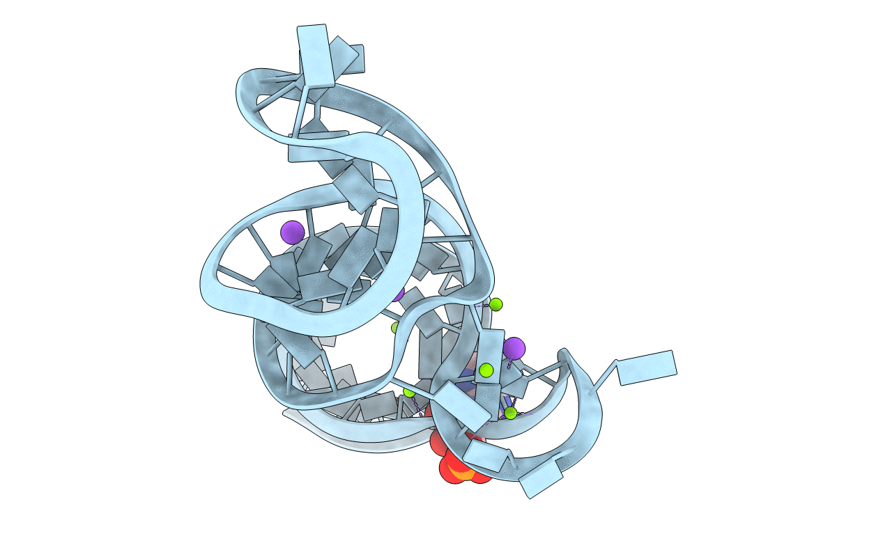
Title:
Crystal structure of the ADP-binding domain of the NAD+ riboswitch with Adenosine monophosphate (AMP)
Biological Source:
Source Organism:
Candidatus Koribacter versatilis Ellin345 (Taxon ID: 204669)
Method Details:
Experimental Method:
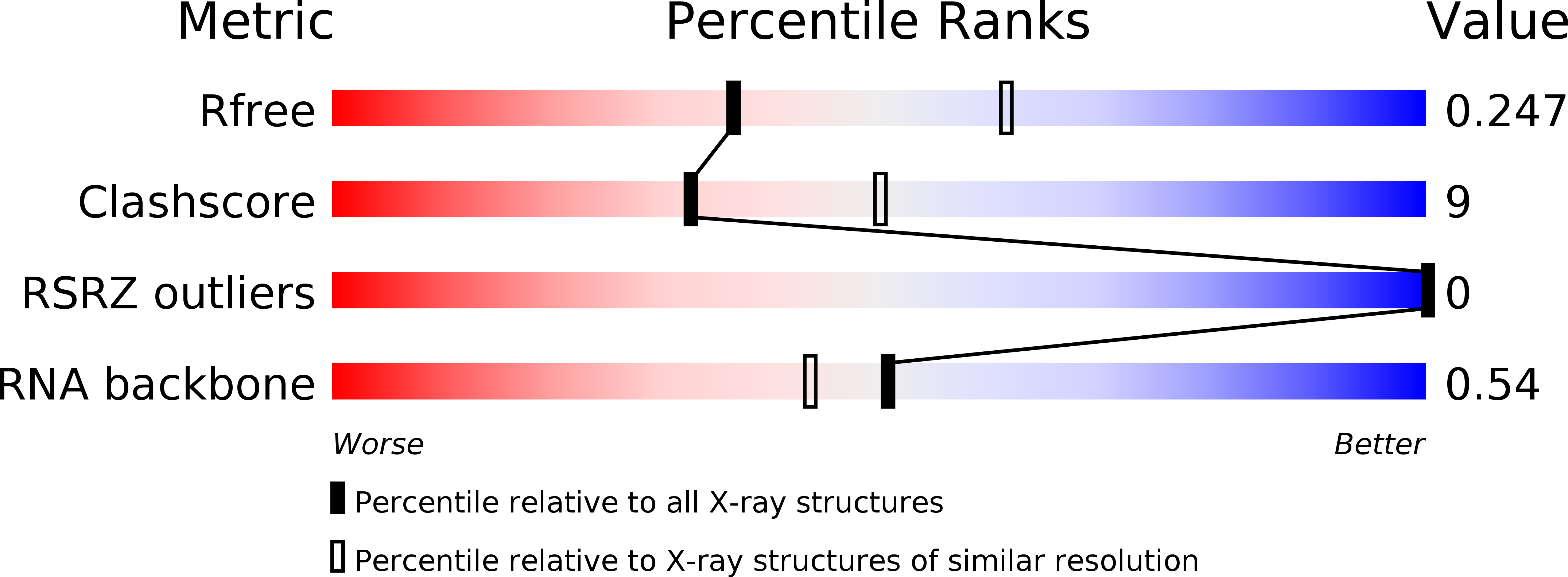
Resolution:
2.53 Å
R-Value Free:
0.23
R-Value Work:
0.20
R-Value Observed:
0.20
Space Group:
I 2 2 2