5YYB image
Entry Detail
PDB ID:
5YYB
Title:
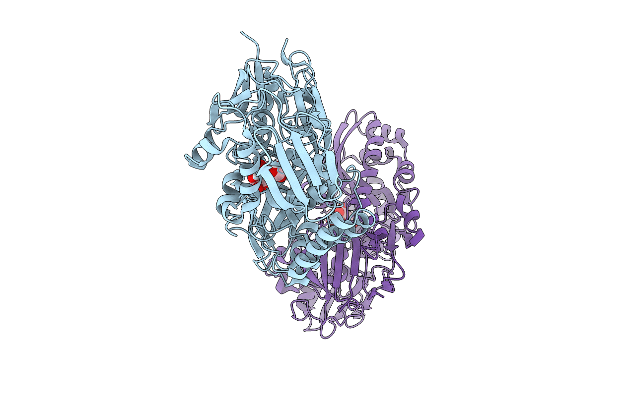
Crystal structure of Sialic acid Binding protein from Haemophilus ducreyi with Neu5Gc
Biological Source:
Host Organism:
PDB Version:
Deposition Date:
2017-12-08
Release Date:
2018-10-24
Method Details:
Experimental Method:
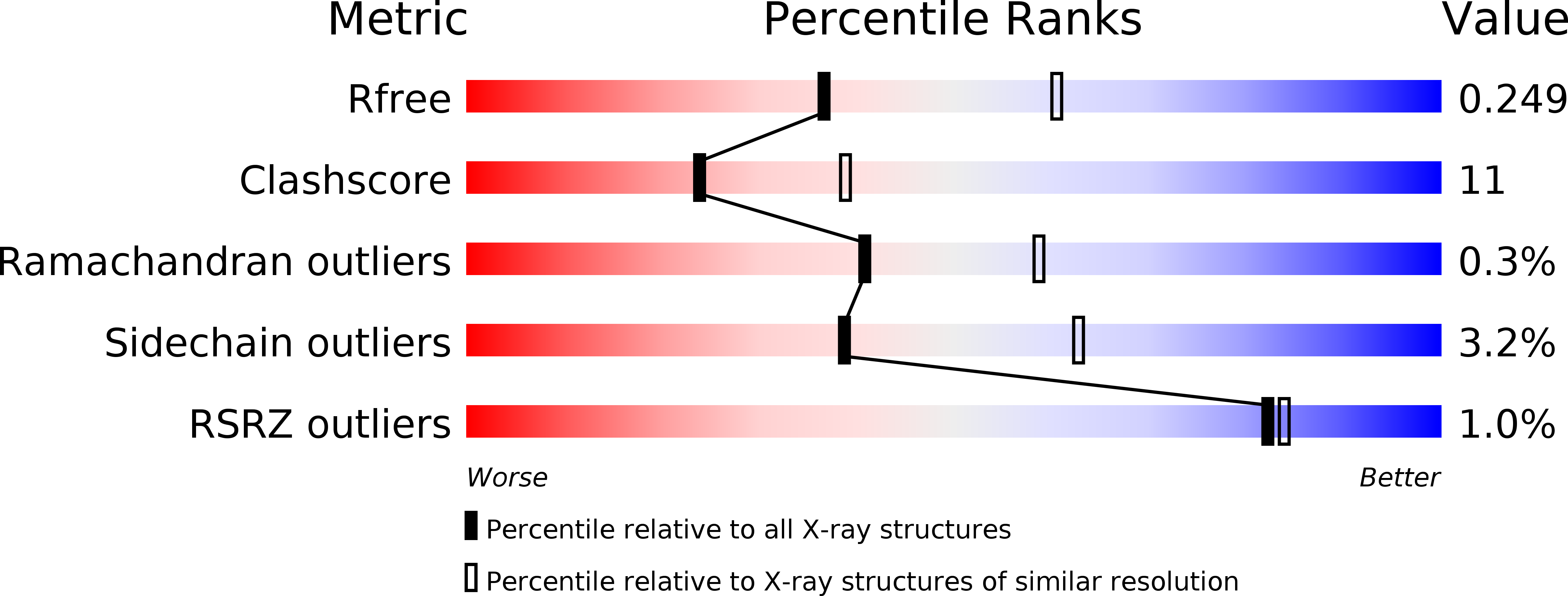
Resolution:
2.48 Å
R-Value Free:
0.24
R-Value Work:
0.17
R-Value Observed:
0.17
Space Group:
C 1 2 1
Macromolecular Entities
Polymer Type:polypeptide(L)
Description:Putative ABC transporter periplasmic binding protein
Chain IDs:A, B
Chain Length:482
Number of Molecules:2
Biological Source:Haemophilus ducreyi 35000HP
Modified Residue
Compound ID Chain ID Parent Comp ID Details 2D Image
MSE A MET modified residue
Ligand Molecules
Primary Citation
Molecular characterization of the interaction of sialic acid with the periplasmic binding protein fromHaemophilus ducreyi.
J. Biol. Chem. 293 20073 20084 (2018)
PMID: 30315109 DOI: 10.1074/jbc.RA118.005151

Abstact

The primary role of bacterial periplasmic binding proteins is sequestration of essential metabolites present at a low concentration in the periplasm and making them available for active transporters that transfer these ligands into the bacterial cell. The periplasmic binding proteins (SiaPs) from the tripartite ATP-independent periplasmic (TRAP) transport system that transports mammalian host-derived sialic acids have been well studied from different pathogenic bacteria, including Haemophilus influenzae, Fusobacterium nucleatum, Pasteurella multocida, and Vibrio cholerae SiaPs bind the sialic acid N-acetylneuraminic acid (Neu5Ac) with nanomolar affinity by forming electrostatic and hydrogen-bonding interactions. Here, we report the crystal structure of a periplasmic binding protein (SatA) of the ATP-binding cassette (ABC) transport system from the pathogenic bacterium Haemophilus ducreyi The structure of Hd-SatA in the native form and sialic acid-bound forms (with Neu5Ac and N-glycolylneuraminic acid (Neu5Gc)), determined to 2.2, 1.5, and 2.5 Å resolutions, respectively, revealed a ligand-binding site that is very different from those of the SiaPs of the TRAP transport system. A structural comparison along with thermodynamic studies suggested that similar affinities are achieved in the two classes of proteins through distinct mechanisms, one enthalpically driven and the other entropically driven. In summary, our structural and thermodynamic characterization of Hd-SatA reveals that it binds sialic acids with nanomolar affinity and that this binding is an entropically driven process. This information is important for future structure-based drug design against this pathogen and related bacteria.

Legend

Protein

Chemical

Disease

Primary Citation of related structures