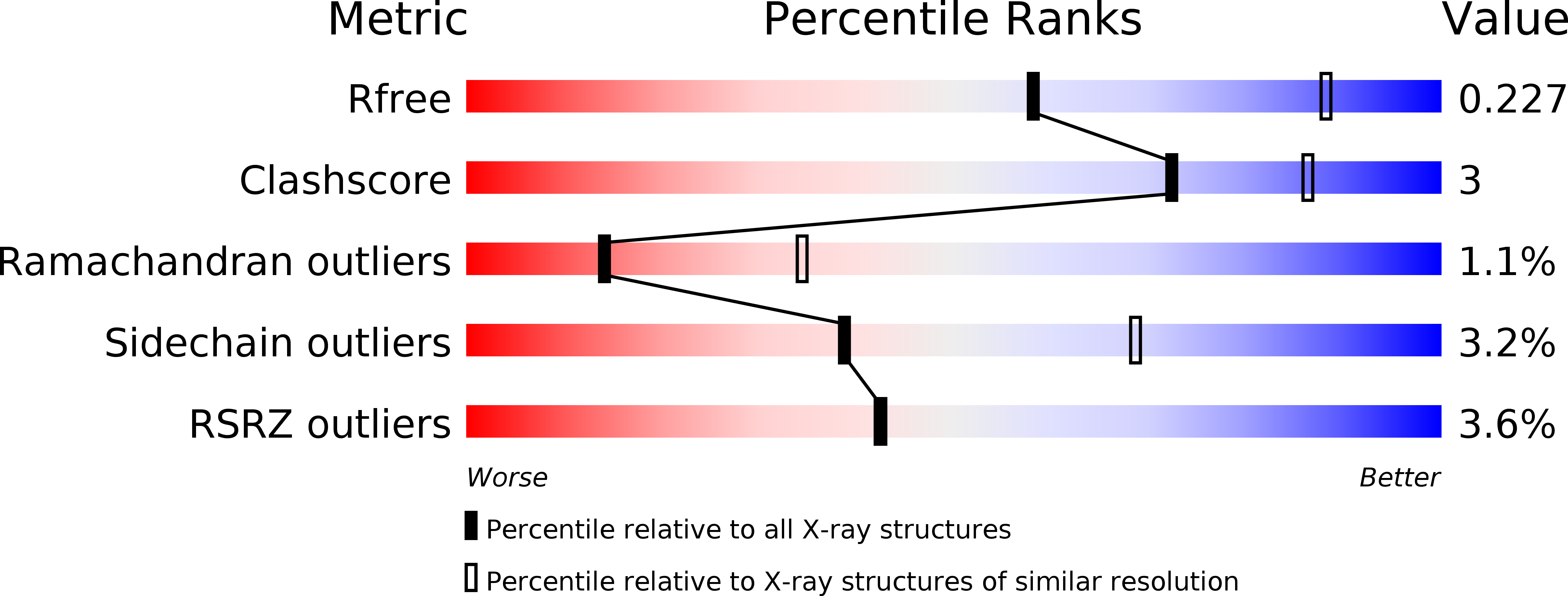
4ZHT image
Deposition Date 2015-04-27
Release Date 2016-06-01
Last Version Date 2024-11-13
Entry Detail
PDB ID:
4ZHT
Keywords:
Title:
Crystal structure of UDP-GlcNAc 2-epimerase
Biological Source:
Source Organism:
Homo sapiens (Taxon ID: 9606)
Host Organism:
Method Details:
Experimental Method:
Resolution:
2.69 Å
R-Value Free:
0.22
R-Value Work:
0.18
R-Value Observed:
0.19
Space Group:
C 1 2 1
Macromolecular Entities
Polymer Type:polypeptide(L)
Molecule:Bifunctional UDP-N-acetylglucosamine 2-epimerase/N-acetylmannosamine kinase
Gene (Uniprot):GNE
Chain IDs:A, B, C, D
Chain Length:411
Number of Molecules:4
Biological Source:Homo sapiens
Modified Residue
Compound ID Chain ID Parent Comp ID Details 2D Image
MSE A MET modified residue
Primary Citation
Mechanism and inhibition of human UDP-GlcNAc 2-epimerase, the key enzyme in sialic acid biosynthesis.
Sci Rep 6 23274 23274 (2016)
PMID: 26980148 DOI: 10.1038/srep23274

Abstact

The bifunctional enzyme UDP-GlcNAc 2-epimerase/ManNAc kinase (GNE) plays a key role in sialic acid production. It is different from the non-hydrolyzing enzymes for bacterial cell wall biosynthesis, and it is feed-back inhibited by the downstream product CMP-Neu5Ac. Here the complex crystal structure of the N-terminal epimerase part of human GNE shows a tetramer in which UDP binds to the active site and CMP-Neu5Ac binds to the dimer-dimer interface. The enzyme is locked in a tightly closed conformation. By comparing the UDP-binding modes of the non-hydrolyzing and hydrolyzing UDP-GlcNAc epimerases, we propose a possible explanation for the mechanistic difference. While the epimerization reactions of both enzymes are similar, Arg113 and Ser302 of GNE are likely involved in product hydrolysis. On the other hand, the CMP-Neu5Ac binding mode clearly elucidates why mutations in Arg263 and Arg266 can cause sialuria. Moreover, full-length modelling suggests a channel for ManNAc trafficking within the bifunctional enzyme.

Legend

Protein

Chemical

Disease

Primary Citation of related structures