Deposition Date
2011-03-21
Release Date
2011-09-07
Last Version Date
2023-12-20
Entry Detail
PDB ID:
2YDH
Keywords:
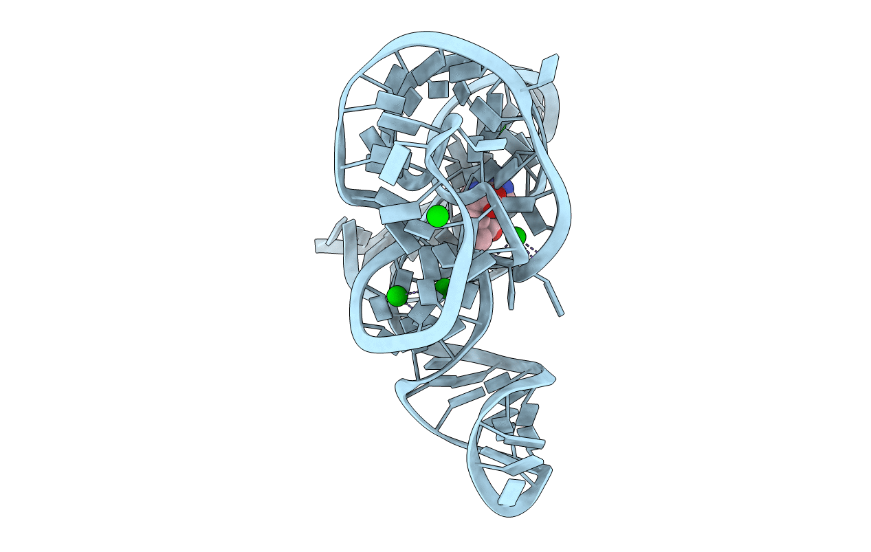
Title:
Crystal structure of the SAM-I riboswitch A94G U34 G18U G19U variant in complex with SAM
Biological Source:
Source Organism(s):
THERMOANAEROBACTER TENGCONGENSIS (Taxon ID: 119072)
Method Details:
Experimental Method:
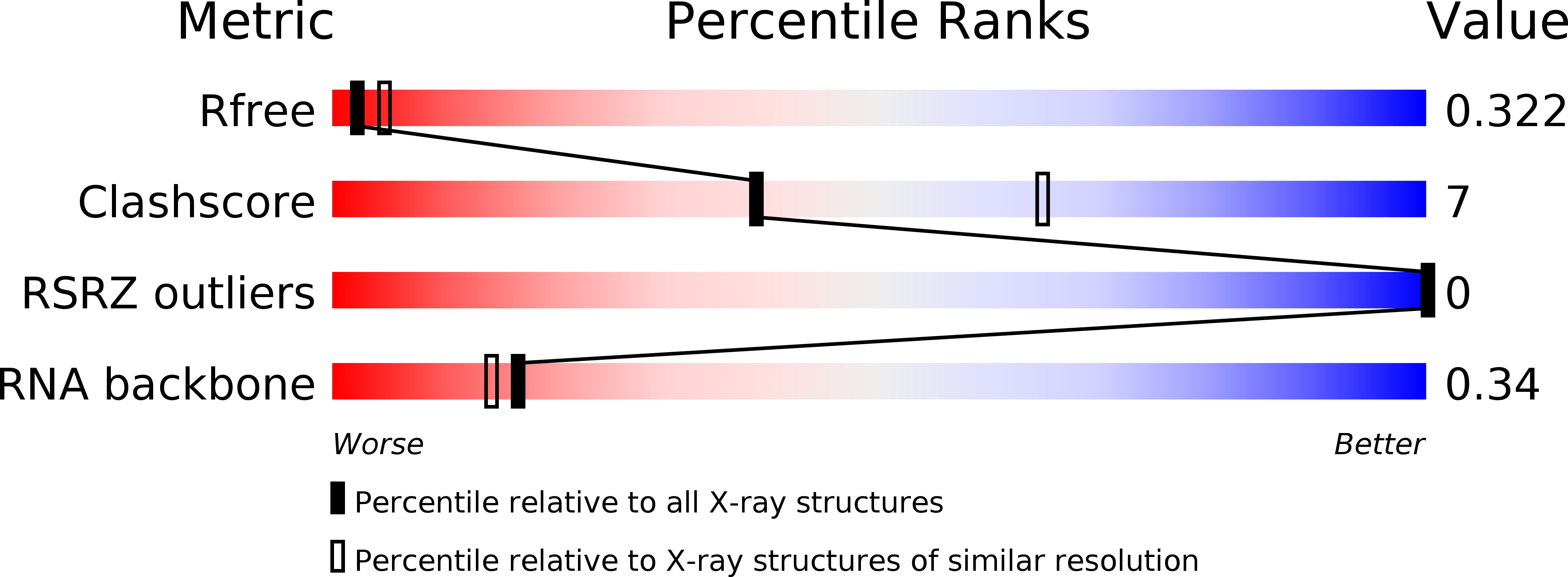
Resolution:
2.90 Å
R-Value Free:
0.32
R-Value Work:
0.26
R-Value Observed:
0.27
Space Group:
P 43 21 2