Deposition Date
2010-04-12
Release Date
2010-08-04
Last Version Date
2023-12-20
Entry Detail
PDB ID:
2XBK
Keywords:
Title:
X-ray structure of the substrate-bound cytochrome P450 PimD - a polyene macrolide antibiotic pimaricin epoxidase
Biological Source:
Source Organism(s):
STREPTOMYCES NATALENSIS (Taxon ID: 68242)
Expression System(s):
Method Details:
Experimental Method:
Resolution:
1.95 Å
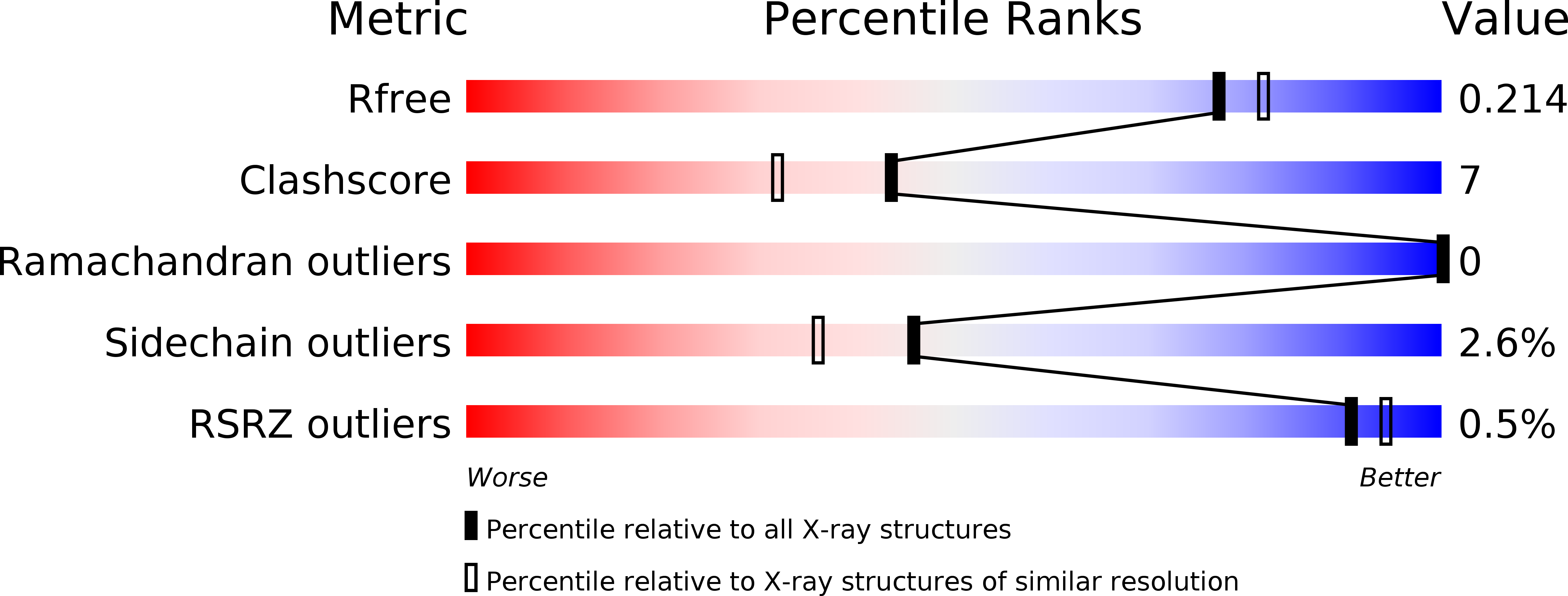
R-Value Free:
0.21
R-Value Work:
0.14
R-Value Observed:
0.15
Space Group:
C 1 2 1