2EUD image
Deposition Date 2005-10-28
Release Date 2006-03-07
Last Version Date 2024-02-14
Entry Detail
PDB ID:
2EUD
Keywords:
Title:
Structures of Yeast Ribonucleotide Reductase I complexed with Ligands and Subunit Peptides
Biological Source:
Source Organism:
Host Organism:
Method Details:
Experimental Method:
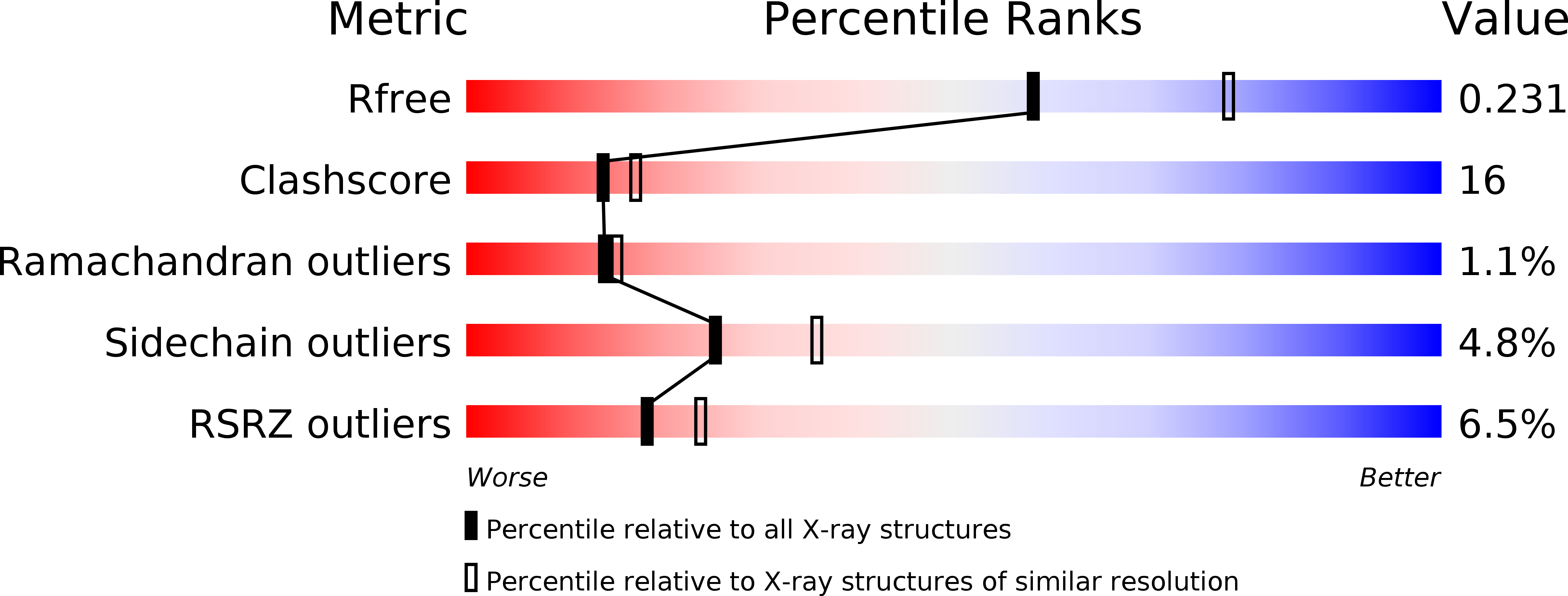
Resolution:
2.30 Å
R-Value Free:
0.24
R-Value Work:
0.20
R-Value Observed:
0.21
Space Group:
P 21 21 2
Macromolecular Entities
Structures with similar UniProt ID
Protein Blast
Polymer Type:polypeptide(L)
Molecule:Ribonucleoside-diphosphate reductase large chain 1
Gene (Uniprot):RNR1
Chain IDs:A
Chain Length:888
Number of Molecules:1
Biological Source:Saccharomyces cerevisiae
Primary Citation
Structures of eukaryotic ribonucleotide reductase I define gemcitabine diphosphate binding and subunit assembly.
Proc.Natl.Acad.Sci.Usa 103 4028 4033 (2006)
PMID: 16537480 DOI: 10.1073/pnas.0600440103

Abstact

Ribonucleotide reductase (RNR) catalyzes the conversion of nucleoside diphosphates to deoxynucleoside diphosphates. Crucial for rapidly dividing cells, RNR is a target for cancer therapy. In eukaryotes, RNR comprises a heterooligomer of alpha(2) and beta(2) subunits. Rnr1, the alpha subunit, contains regulatory and catalytic sites; Rnr2, the beta subunit (in yeast, a heterodimer of Rnr2 and Rnr4), houses the diferric-tyrosyl radical crucial for catalysis. Here, we present three x-ray structures of eukaryotic Rnr1 from Saccharomyces cerevisiae: one bound to gemcitabine diphosphate (GemdP), the active metabolite of the mechanism-based chemotherapeutic agent gemcitabine; one with an Rnr2-derived peptide, and one with an Rnr4-derived peptide. Our structures reveal that GemdP binds differently from its analogue, cytidine diphosphate; because of unusual interactions of the geminal fluorines, the ribose and base of GemdP shift substantially, and loop 2, which mediates substrate specificity, adopts different conformations when binding to GemdP and cytidine diphosphate. The Rnr2 and Rnr4 peptides, which block RNR assembly, bind differently from each other but have unique modes of binding not seen in prokaryotic RNR. The Rnr2 peptide adopts a conformation similar to that previously reported from an NMR study for a mouse Rnr2-based peptide. In yeast, the Rnr2 peptide binds at subsites consisting of residues that are highly conserved among yeast, mouse, and human Rnr1s, suggesting that the mode of Rnr1-Rnr2 binding is conserved among eukaryotes. These structures provide new insights into subunit assembly and a framework for structure-based drug design targeting RNR.

Legend

Protein

Chemical

Disease

Primary Citation of related structures
Feedback Form
Name
Email
Institute
Feedback