1VQ2 image
Deposition Date 2004-12-15
Release Date 2004-12-21
Last Version Date 2023-12-27
Entry Detail
PDB ID:
1VQ2
Keywords:
Title:
CRYSTAL STRUCTURE OF T4-BACTERIOPHAGE DEOXYCYTIDYLATE DEAMINASE, MUTANT R115E
Biological Source:
Source Organism:
Host Organism:
Method Details:
Experimental Method:
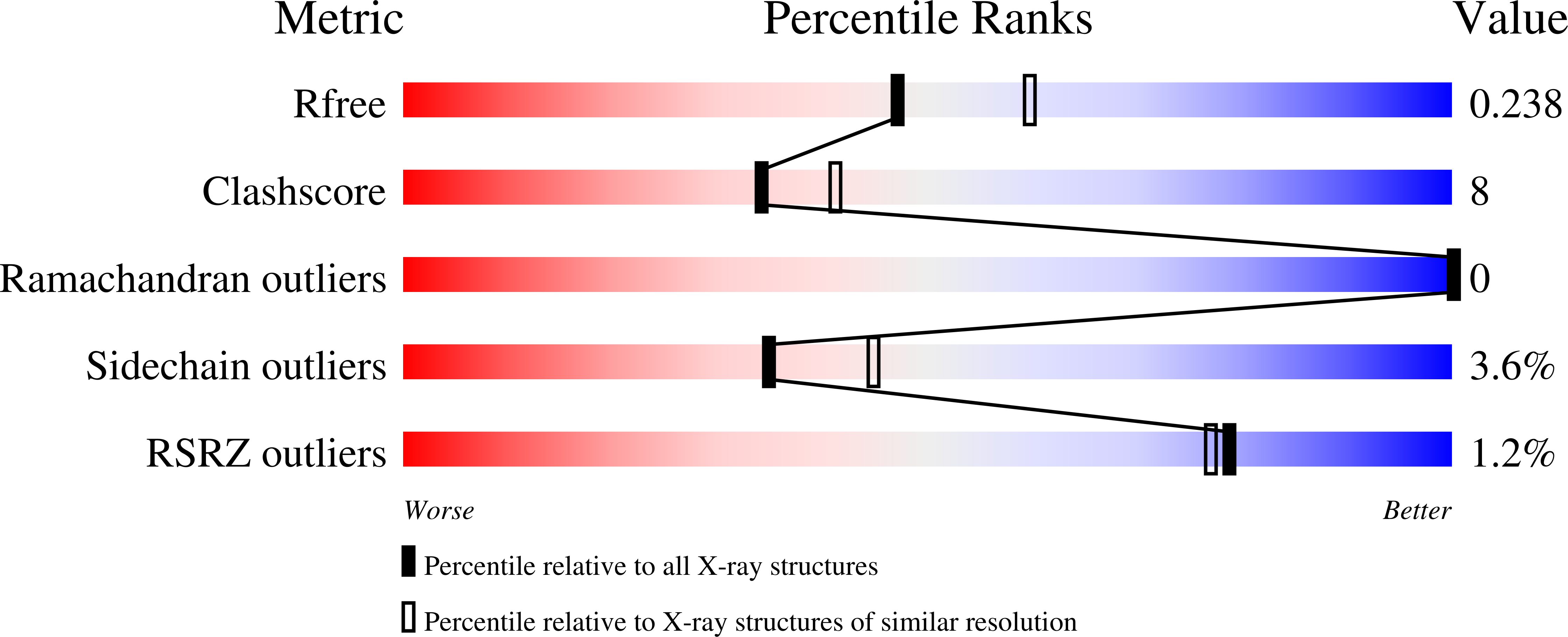
Resolution:
2.20 Å
R-Value Free:
0.24
R-Value Work:
0.21
R-Value Observed:
0.21
Space Group:
P 63 2 2
Macromolecular Entities
Polymer Type:polypeptide(L)
Molecule:DEOXYCYTIDYLATE DEAMINASE
Gene (Uniprot):CD
Mutations:R115E
Chain IDs:A
Chain Length:193
Number of Molecules:1
Biological Source:Enterobacteria phage T4
Primary Citation
Three-Dimensional Structure of the R115E Mutant of T4-Bacteriophage 2'-Deoxycytidylate Deaminase
Biochemistry 43 13715 ? (2004)
PMID: 15504034 DOI: 10.1021/bi048928h

Abstact

2'-Deoxycytidylate deaminase (dCD) converts deoxycytidine 5'-monophosphate (dCMP) to deoxyuridine 5'-monophosphate and is a major supplier of the substrate for thymidylate synthase, an important enzyme in DNA synthesis and a major target for cancer chemotherapy. Wild-type dCD is allosterically regulated by the end products of its metabolic pathway, deoxycytidine 5'-triphosphate and deoxythymidine 5'-triphosphate, which act as an activator and an inhibitor, respectively. The first crystal structure of a dCD, in the form of the R115E mutant of the T4-bacteriophage enzyme complexed with the active site inhibitor pyrimidin-2-one deoxyribotide, has been determined at 2.2 A resolution. This mutant of dCD is active, even in the absence of the allosteric regulators. The molecular topology of dCD is related to that of cytidine deaminase (CDA) but with modifications for formation of the binding site for the phosphate group of dCMP. The enzyme has a zinc ion-based mechanism that is similar to that of CDA. A second zinc ion that is present in bacteriophage dCD, but absent in mammalian dCD and CDA, is important for the structural integrity of the enzyme and for the binding of the phosphate group of the substrate or inhibitor. Although the R115E mutant of dCD is a dimer in solution, it crystallizes as a hexamer, mimicking the natural state of the wild-type enzyme. Residues 112 and 115, which are known to be important for the binding of the allosteric regulators, are found in a pocket that is at the intersubunit interfaces in the hexamer but distant from the substrate-binding site. The substrate-binding site is composed of residues from a single protein molecule and is sequestered in a deep groove. This groove is located at the outer surface of the hexamer but ends at the subunit interface that also includes residue 115. It is proposed that the absence of subunit interactions at this interface in the dimeric R115E mutant renders the substrate-binding site accessible. In contrast, for the wild-type enzyme, binding of dCTP induces an allosteric effect that affects the subunit interactions and results in an increase in the accessibility of the binding site.

Legend

Protein

Chemical

Disease

Primary Citation of related structures