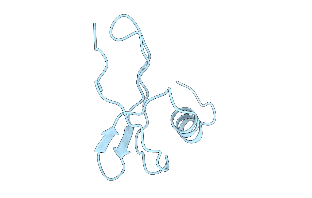
1IKL image
Deposition Date 1995-08-03
Release Date 1995-10-15
Last Version Date 2024-11-13
Entry Detail
PDB ID:
1IKL
Title:
NMR study of monomeric human interleukin-8 (minimized average structure)
Biological Source:
Source Organism:
Homo sapiens (Taxon ID: 9606)
Method Details:
Experimental Method:
Conformers Submitted:
1
Macromolecular Entities
Polymer Type:polypeptide(L)
Molecule:HUMAN INTERLEUKIN-8 (MONOMERIC)
Gene (Uniprot):CXCL8
Mutations:DEL(1-3), H LEU 25 MODIFIED TO N-METHYL
Chain IDs:A
Chain Length:72
Number of Molecules:1
Biological Source:Homo sapiens
Primary Citation
1H NMR solution structure of an active monomeric interleukin-8.
Biochemistry 34 12983 12990 (1995)
PMID: 7548056 DOI: 10.1021/bi00040a008

Abstact

The solution structure of a monomeric form of interleukin-8 (IL-8) has been solved using 1H NMR spectroscopy. The chemically synthesized nonnatural analog [IL-8 (4-72) L25 NH-->NCH3] has the same activity as that of native IL-8. Thirty structures were generated using the hybrid distance geometry and simulated annealing protocol using the program X-PLOR. The structure is well-defined except for N-terminal residues 4-6 and C-terminal residues 67-72. The rms distribution about the average structure for residues 7-66 is 0.38 A for the backbone atoms and 0.87 A for all heavy atoms. The structure consists of a series of turns and loops followed by a triple-stranded beta sheet and a C-terminal alpha helix. The structure of the monomer is largely similar to the native dimeric IL-8 structures previously determined by both NMR and X-ray methods. The major difference is that, in the monomeric analog, the C-terminal residues 67-72 are disordered whereas they are helical in the two dimeric structures. The best fit superposition of the backbone atoms of residues 7-66 of the monomer structure on the dimeric IL-8 structures showed rms differences of 1.5 and 1.2 A respectively. The turn (residues 31-35), which is disulfide linked to the N-terminal region, adopts a conformation in the monomer similar to that seen in the dimeric X-ray structure (rms difference 1.4 A) and different from that seen in the dimeric NMR structure (rms difference 2.7 A).(ABSTRACT TRUNCATED AT 250 WORDS)

Legend

Protein

Chemical

Disease

Primary Citation of related structures