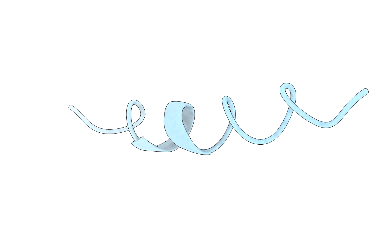
1AWY image
Entry Detail
PDB ID:
1AWY
Keywords:
Title:
NMR STRUCTURE OF CALCIUM BOUND CONFORMER OF CONANTOKIN G, MINIMIZED AVERAGE STRUCTURE
Biological Source:
Source Organism:
PDB Version:
Deposition Date:
1997-10-06
Release Date:
1998-04-08
Method Details:
Experimental Method:
Conformers Calculated:
25
Conformers Submitted:
1
Selection Criteria:
LOWEST ENERGY
Macromolecular Entities
Polymer Type:polypeptide(L)
Description:CONANTOXIN G
Chain IDs:A
Chain Length:18
Number of Molecules:1
Biological Source:Conus geographus
Modified Residue
Compound ID Chain ID Parent Comp ID Details 2D Image
CGU A GLU GAMMA-CARBOXY-GLUTAMIC ACID
Ligand Molecules
Primary Citation
Role of gamma-carboxyglutamic acid in the calcium-induced structural transition of conantokin G, a conotoxin from the marine snail Conus geographus.
Biochemistry 36 15677 15684 (1997)
PMID: 9398296 DOI: 10.1021/bi9718550

Abstact

Conantokin G is a gamma-carboxyglutamic acid- (Gla-) containing conotoxin isolated from the venom of the marine cone snail Conus geographus. This 17-residue polypeptide, which contains five gamma-carboxyglutamic acid residues, is a N-methyl-d-aspartate- (NMDA-) type glutamate receptor antagonist. To investigate the role of gamma-carboxyglutamic acid in the calcium-induced structural transition of conantokin G, we determined the three-dimensional structure of the conantokin G/Ca2+ complex by two-dimensional 1H NMR spectroscopy and compared it to the high-resolution structure of conantokin G in the absence of metal ions [Rigby et al. (1997) Biochemistry 36, 6906]. Complete resonance assignments were made by two dimensional 1H NMR spectroscopy at pH 5.6 in the presence of saturating amounts of Ca2+. Distance geometry and simulated annealing methods were used to derive 23 convergent structures from a set of 302 interproton distance restraints and two torsion angle measurements. A high-resolution structure, with the backbone root mean square deviation to the geometric average of the 23 structures of 0.6 +/- 0.1 A, contains a linear alpha-helix from Gla 3 to Lys 15. Gla residues 3, 7, 10, and 14 are aligned in a linear array on one face of the helix. A genetic algorithm was applied to determine the calcium positions in conantokin G, and the conantokin G/Ca2+ complex refined by molecular simulation. Upon binding of Ca2+ to gamma-carboxyglutamic acid, conantokin G undergoes a conformational transition from a distorted curvilinear 310 helix to a linear alpha-helix. Occupancy of the metal binding sites, defined by gamma-carboxyglutamic acids, results in formation of a calcium-carboxylate network that linearizes the helix and exposes the hydrophobic amino acids on the opposite face of the helix.

Legend

Protein

Chemical

Disease

Primary Citation of related structures