
Search Count: 15
 |
Organism: Homo sapiens
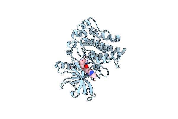
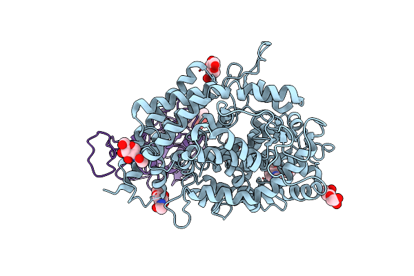
Method: X-RAY DIFFRACTION Resolution:1.60 Å Release Date: 2025-06-25 Classification: CELL CYCLE Ligands: A1A6M, CL, MG |
 |
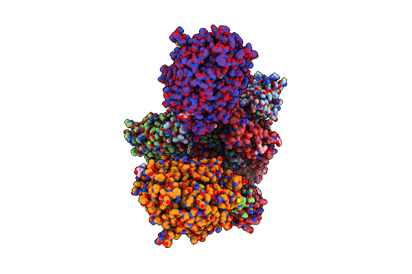
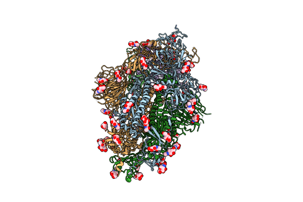
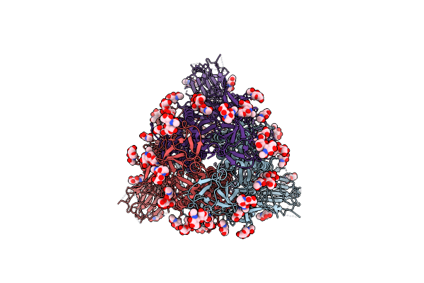
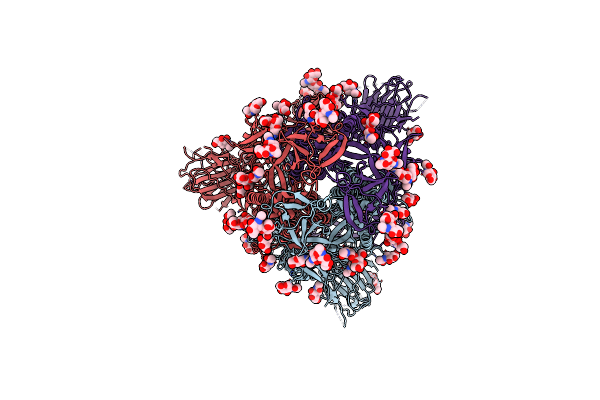
Organism: Severe acute respiratory syndrome coronavirus 2, Homo sapiens
Method: ELECTRON MICROSCOPY Release Date: 2020-12-16 Classification: HYDROLASE/VIRAL PROTEIN Ligands: NAG |
 |
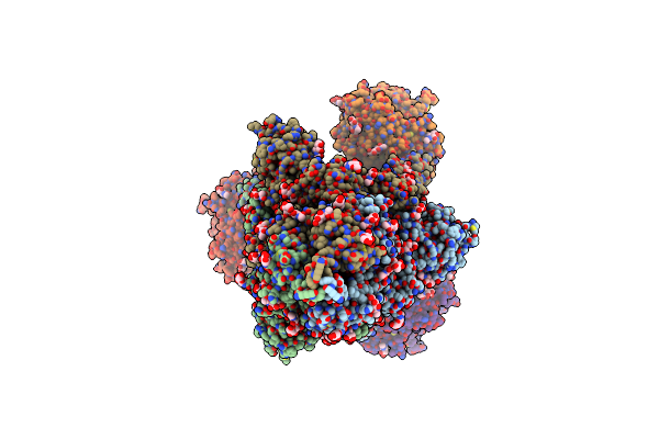
Organism: Severe acute respiratory syndrome coronavirus 2, Homo sapiens
Method: ELECTRON MICROSCOPY Release Date: 2020-12-16 Classification: HYDROLASE/VIRAL PROTEIN Ligands: NAG |
 |
Organism: Severe acute respiratory syndrome coronavirus 2, Homo sapiens
Method: ELECTRON MICROSCOPY Release Date: 2020-12-16 Classification: VIRAL PROTEIN/Hydrolase Ligands: NAG |
 |
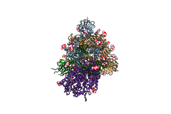
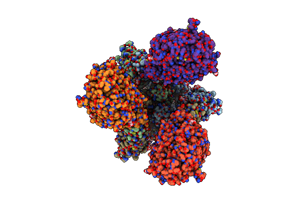
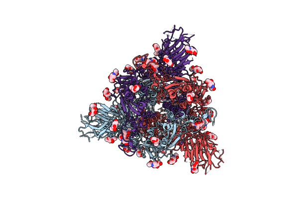
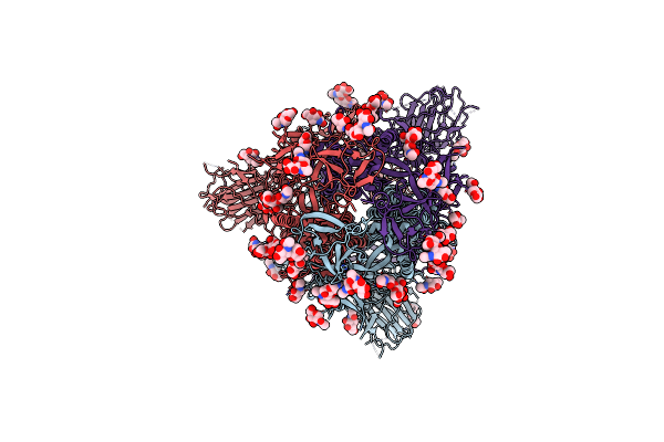
Ace2-Rbd Focused Refinement Using Symmetry Expansion Of Applied C3 For Triple Ace2-Bound Sars-Cov-2 Trimer Spike At Ph 7.4
Organism: Homo sapiens, Severe acute respiratory syndrome coronavirus 2
Method: ELECTRON MICROSCOPY Release Date: 2020-12-09 Classification: Hydrolase/Viral Protein Ligands: NAG |
 |
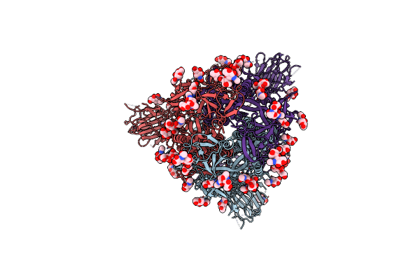
Organism: Homo sapiens, Severe acute respiratory syndrome coronavirus 2
Method: ELECTRON MICROSCOPY Release Date: 2020-12-09 Classification: HYDROLASE/VIRAL PROTEIN Ligands: NAG |
 |
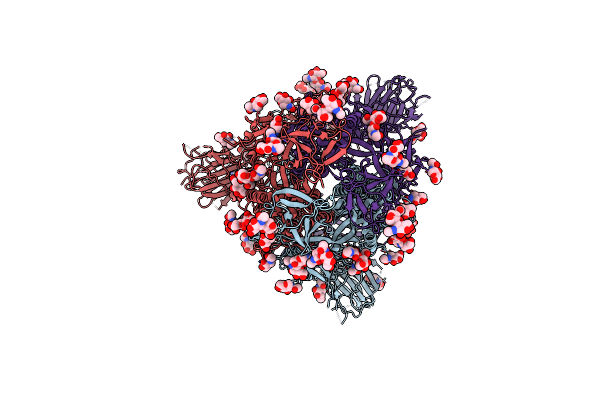
Organism: Severe acute respiratory syndrome coronavirus 2, Homo sapiens
Method: ELECTRON MICROSCOPY Release Date: 2020-12-09 Classification: HYDROLASE/VIRAL PROTEIN Ligands: NAG |
 |
Organism: Severe acute respiratory syndrome coronavirus 2, Homo sapiens
Method: ELECTRON MICROSCOPY Release Date: 2020-12-09 Classification: HYDROLASE/VIRAL PROTEIN Ligands: NAG |
 |
Organism: Severe acute respiratory syndrome coronavirus 2
Method: ELECTRON MICROSCOPY Release Date: 2020-11-25 Classification: VIRAL PROTEIN Ligands: NAG |
 |
Organism: Severe acute respiratory syndrome coronavirus 2
Method: ELECTRON MICROSCOPY Release Date: 2020-08-12 Classification: VIRAL PROTEIN Ligands: NAG |
 |
Organism: Severe acute respiratory syndrome coronavirus 2
Method: ELECTRON MICROSCOPY Release Date: 2020-08-12 Classification: VIRAL PROTEIN Ligands: NAG |
 |
Organism: Severe acute respiratory syndrome coronavirus 2
Method: ELECTRON MICROSCOPY Release Date: 2020-08-12 Classification: VIRAL PROTEIN Ligands: NAG |
 |
Organism: Severe acute respiratory syndrome coronavirus 2
Method: ELECTRON MICROSCOPY Release Date: 2020-08-12 Classification: VIRAL PROTEIN Ligands: NAG |
 |
Organism: Severe acute respiratory syndrome coronavirus 2
Method: ELECTRON MICROSCOPY Release Date: 2020-07-29 Classification: VIRAL PROTEIN Ligands: NAG |
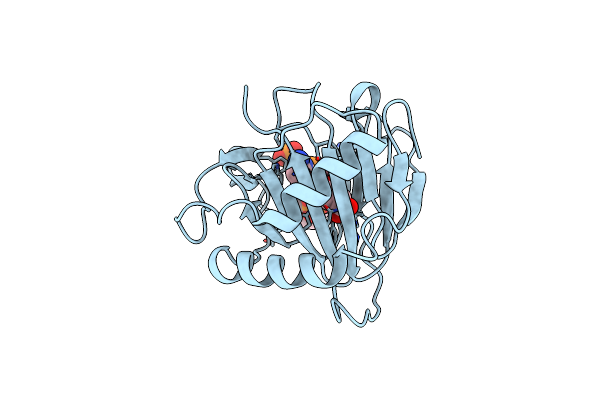 |
Crystal Structure Of Alkb T208A Mutant Protein In Complex With Co(Ii), 2-Oxoglutarate, And Methylated Trinucleotide T-Mea-T
Organism: Escherichia coli (strain k12), Synthetic construct
Method: X-RAY DIFFRACTION Resolution:1.33 Å Release Date: 2015-12-23 Classification: Oxidoreductase/DNA Ligands: CO, AKG |