
Search Count: 61
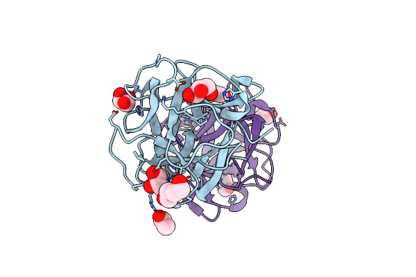 |
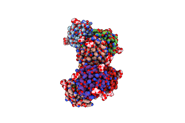
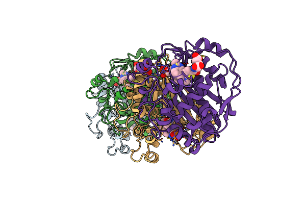
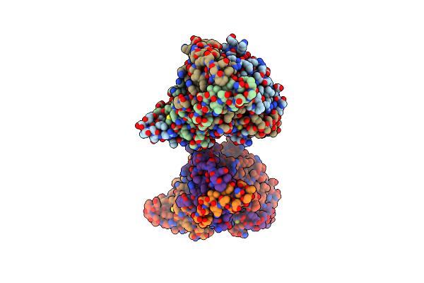
Organism: Mytilus californianus
Method: X-RAY DIFFRACTION Resolution:1.49 Å Release Date: 2024-12-18 Classification: SUGAR BINDING PROTEIN Ligands: GOL, ACT, PEG |
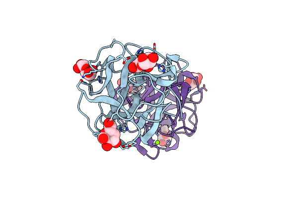 |
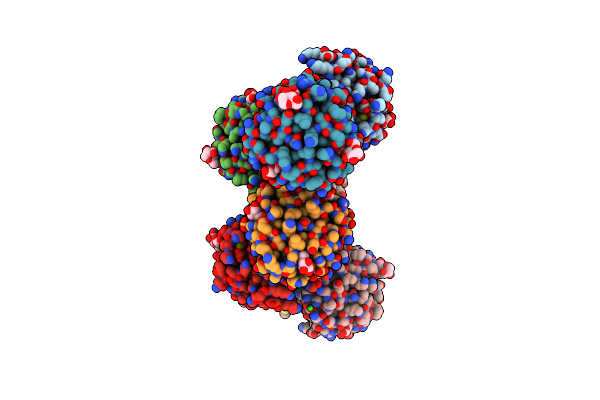
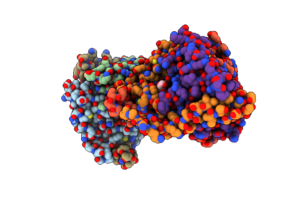
Organism: Mytilus californianus
Method: X-RAY DIFFRACTION Resolution:2.21 Å Release Date: 2024-12-18 Classification: SUGAR BINDING PROTEIN Ligands: GLA, MG |
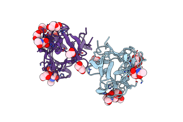 |
Organism: Mytilus californianus
Method: X-RAY DIFFRACTION Resolution:1.66 Å Release Date: 2024-12-18 Classification: SUGAR BINDING PROTEIN Ligands: A2G, GOL, A1AAJ, ACT, PEG |
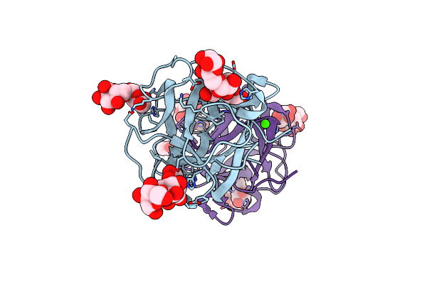 |
Organism: Mytilus californianus
Method: X-RAY DIFFRACTION Resolution:1.58 Å Release Date: 2024-12-18 Classification: SUGAR BINDING PROTEIN Ligands: CA, ACT, GOL |
 |
Organism: Mytilus californianus
Method: X-RAY DIFFRACTION Resolution:2.09 Å Release Date: 2024-12-18 Classification: SUGAR BINDING PROTEIN Ligands: GLA, EPE, PG4, CA, ACT, GOL |
 |
Organism: Mytilus californianus
Method: X-RAY DIFFRACTION Resolution:1.89 Å Release Date: 2024-12-18 Classification: SUGAR BINDING PROTEIN Ligands: GAL, CA |
 |
Organism: Mytilus californianus
Method: X-RAY DIFFRACTION Resolution:1.77 Å Release Date: 2024-12-18 Classification: SUGAR BINDING PROTEIN Ligands: GOL, ACT, PEG, NI |
 |
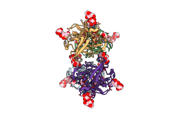
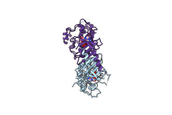
Organism: White spot syndrome virus
Method: X-RAY DIFFRACTION Resolution:2.28 Å Release Date: 2023-06-07 Classification: TRANSFERASE Ligands: UMP, 1PE, SO4, BTB |
 |
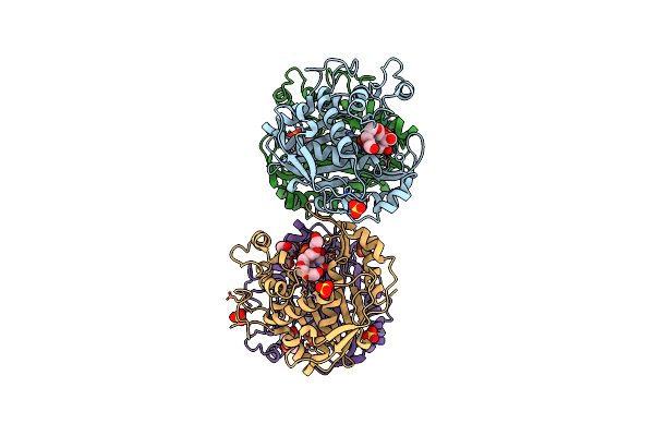
Structure Of Wssv Thymidylate Synthase In Complex With Dump And Raltitrexed
Organism: Shrimp white spot syndrome virus (isolate tongan)
Method: X-RAY DIFFRACTION Resolution:1.43 Å Release Date: 2023-06-07 Classification: TRANSFERASE Ligands: UMP, D16, SO4 |
 |
Organism: Cydia pomonella granulosis virus (isolate mexican)
Method: X-RAY DIFFRACTION Resolution:2.01 Å Release Date: 2021-02-03 Classification: VIRAL PROTEIN Ligands: AMP |
 |
Crystal Structure Of White Spot Syndrome Virus Thymidylate Synthase - Apo Form
Organism: White spot syndrome virus (isolate shrimp/china/tongan/1996)
Method: X-RAY DIFFRACTION Resolution:2.30 Å Release Date: 2020-11-25 Classification: TRANSFERASE Ligands: SO4, PGE |
 |
Crystal Structure Of White Spot Syndrome Virus Thymidylate Synthase - Ternary Complex With Methotrexate And Dump
Organism: White spot syndrome virus (isolate shrimp/china/tongan/1996)
Method: X-RAY DIFFRACTION Resolution:2.75 Å Release Date: 2020-11-25 Classification: TRANSFERASE Ligands: UMP, MTX |
 |
Organism: Drosophila x virus (isolate chung/1996)
Method: X-RAY DIFFRACTION Resolution:2.00 Å Release Date: 2020-11-18 Classification: VIRAL PROTEIN Ligands: SO4 |
 |
Organism: Drosophila x virus (isolate chung/1996)
Method: X-RAY DIFFRACTION Resolution:1.98 Å Release Date: 2020-11-18 Classification: VIRAL PROTEIN Ligands: IMD, GOL |
 |
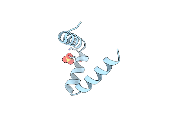
Organism: Sheeppox virus (strain turkey/tu-v02127), Homo sapiens
Method: X-RAY DIFFRACTION Resolution:2.05 Å Release Date: 2020-07-29 Classification: APOPTOSIS |
 |
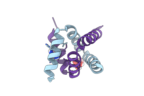
Organism: Sheeppox virus (strain turkey/tu-v02127), Homo sapiens
Method: X-RAY DIFFRACTION Resolution:2.91 Å Release Date: 2020-07-29 Classification: APOPTOSIS |
 |
Organism: Cydia pomonella granulosis virus (isolate mexico/1963)
Method: ELECTRON CRYSTALLOGRAPHY Resolution:1.60 Å Release Date: 2020-04-29 Classification: VIRAL PROTEIN |
 |
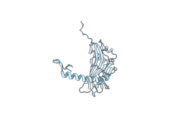
Organism: Cydia pomonella granulosis virus
Method: X-RAY DIFFRACTION Resolution:1.65 Å Release Date: 2019-02-20 Classification: VIRAL PROTEIN Ligands: GOL, CA |
 |
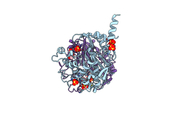
Crystal Structure Of The Dutpase Of White Spot Syndrome Virus In The Apo State
Organism: White spot syndrome virus (isolate shrimp/china/tongan/1996)
Method: X-RAY DIFFRACTION Resolution:2.40 Å Release Date: 2017-12-06 Classification: VIRAL PROTEIN |
 |
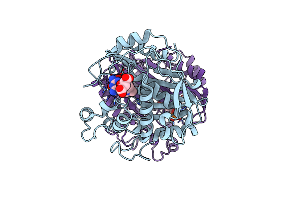
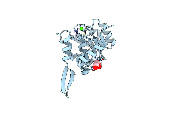
Crystal Structure Of The Dutpase Of White Spot Syndrome Virus In Complex With Du,Ppi And Mg2+
Organism: White spot syndrome virus (isolate shrimp/china/tongan/1996)
Method: X-RAY DIFFRACTION Resolution:2.03 Å Release Date: 2017-12-06 Classification: VIRAL PROTEIN Ligands: DUR, POP, MG |