| Project Accession: | IBIAP_1000000027 |
| Title: | PCSeg: Color model driven probabilistic multiphase level set based tool for plasma cell segmentation in multiple myeloma |
| Representative Image: |  |
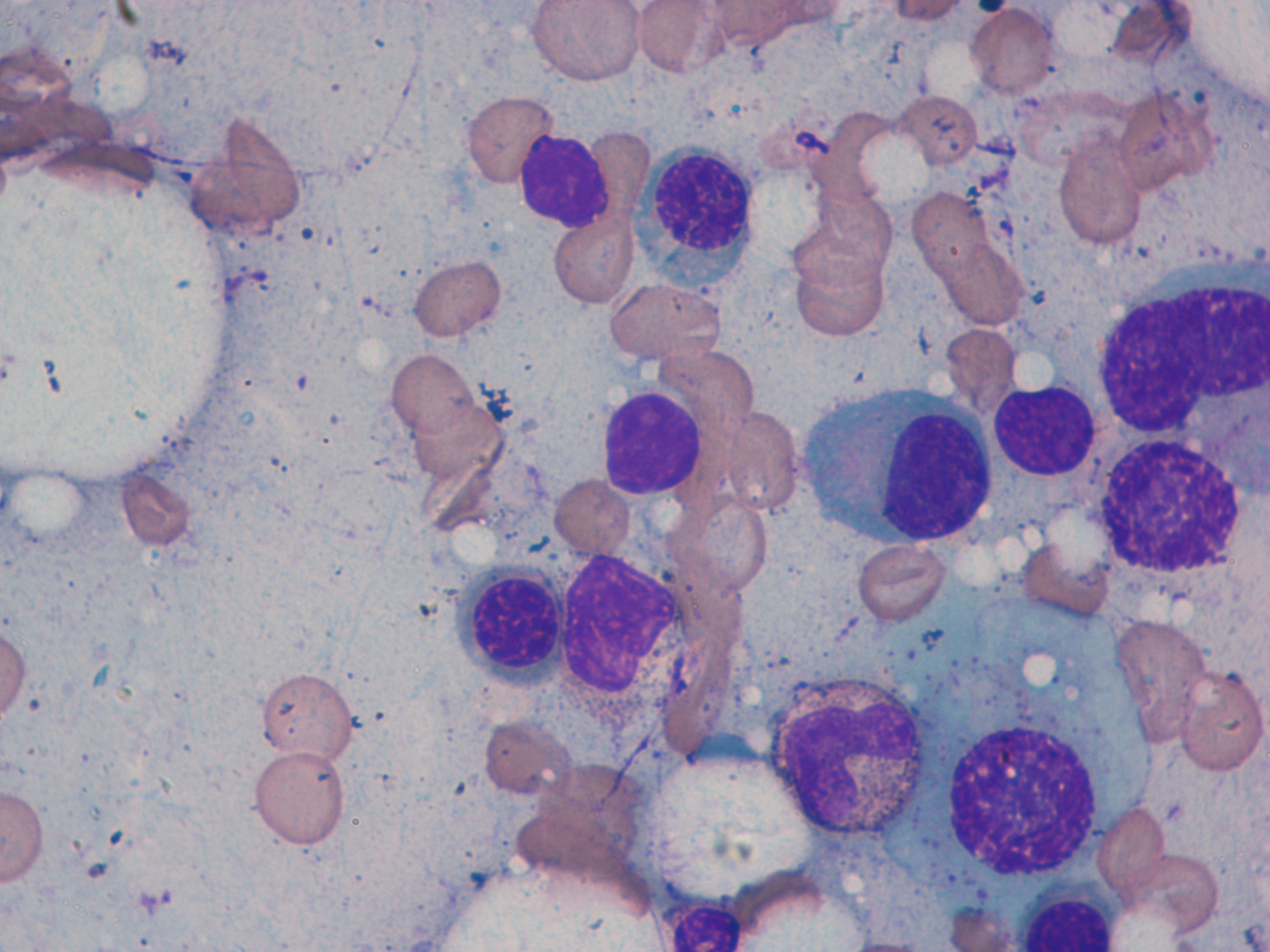
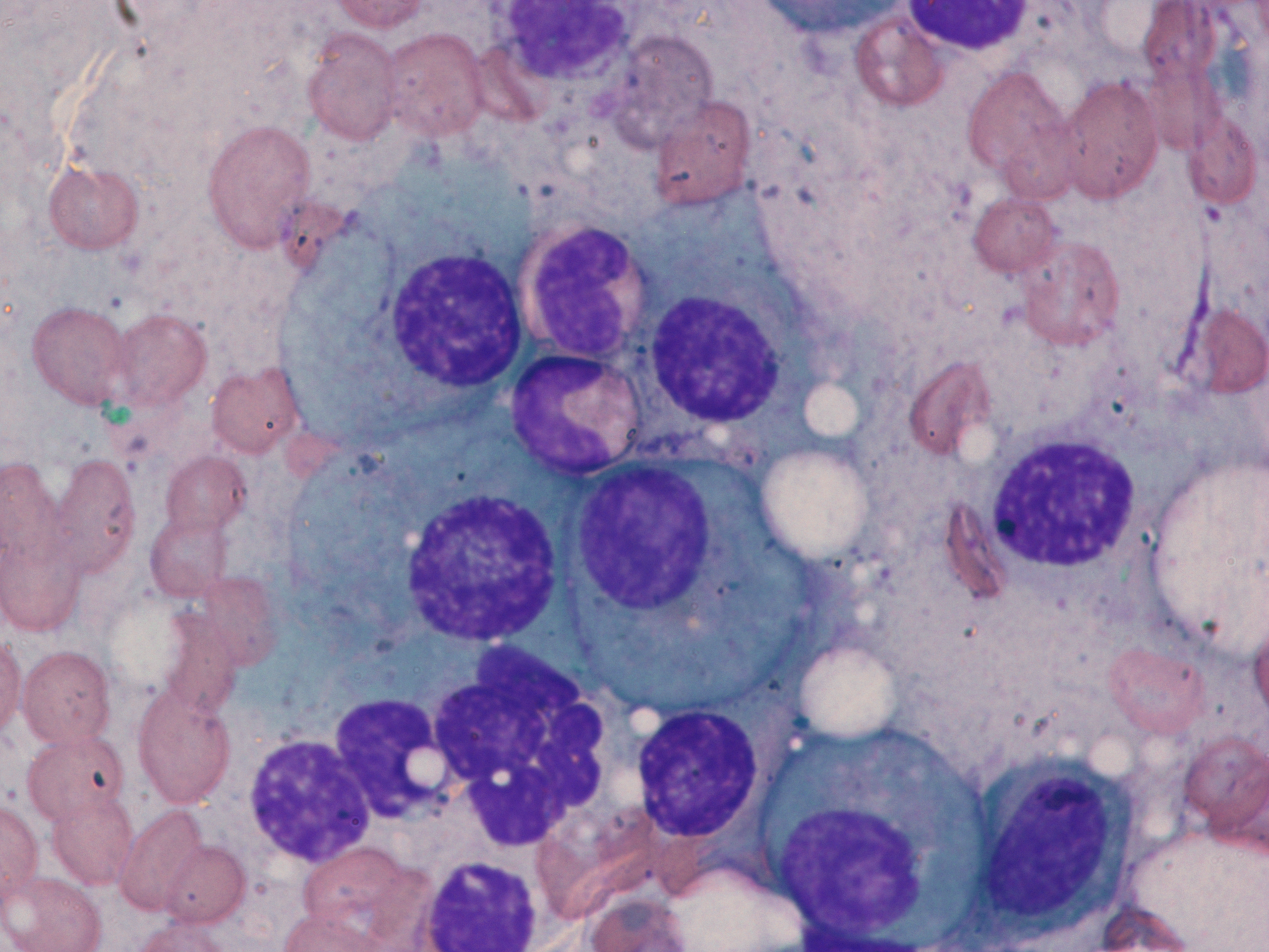
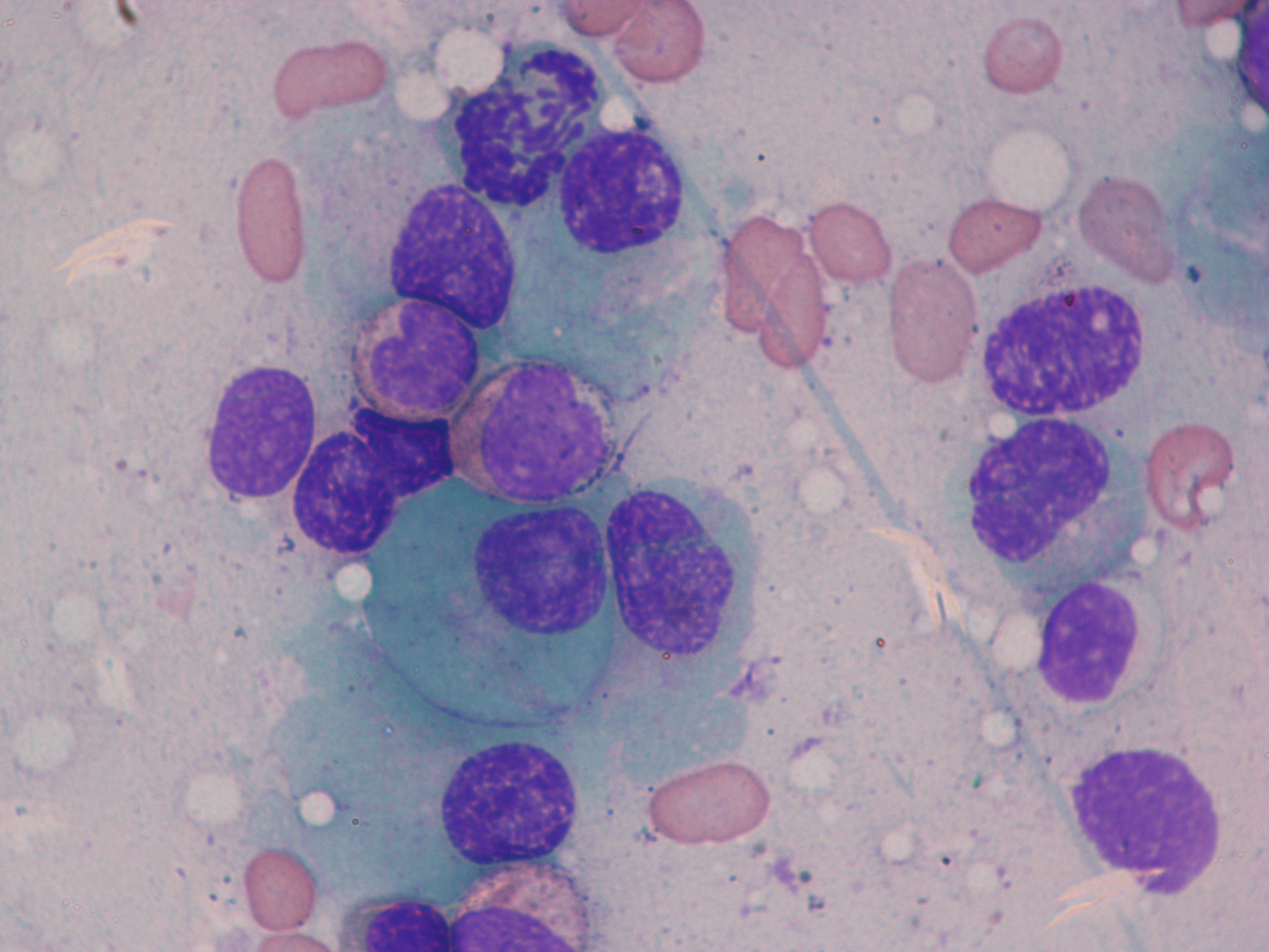
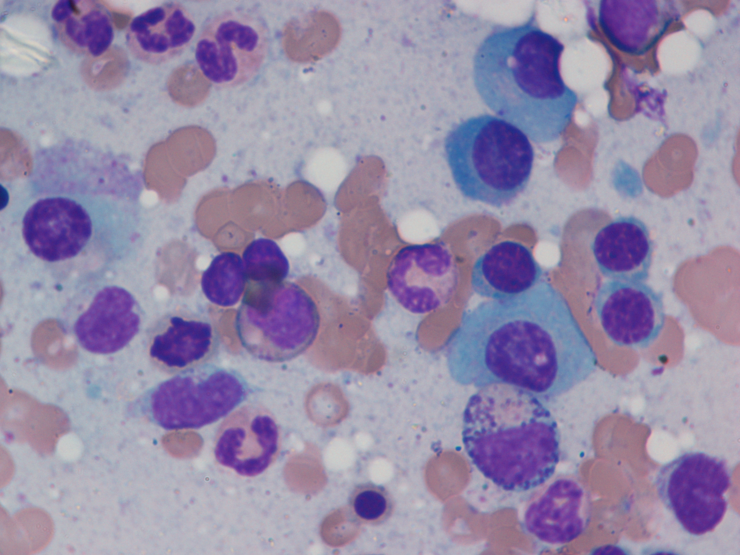
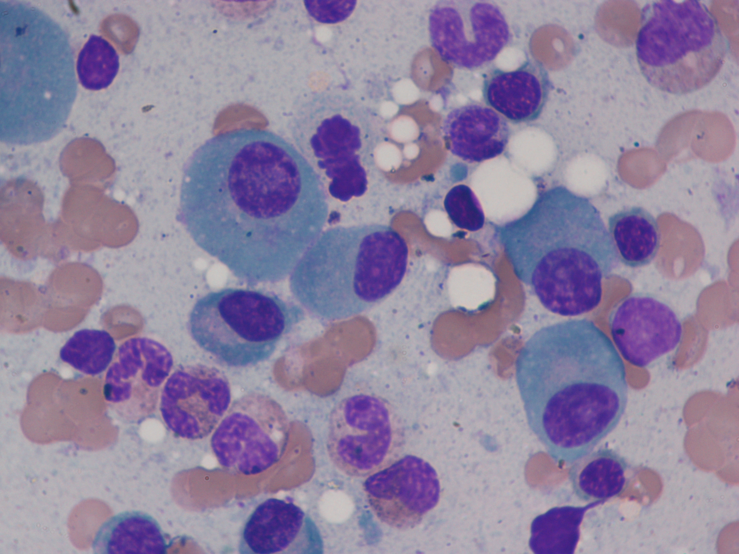
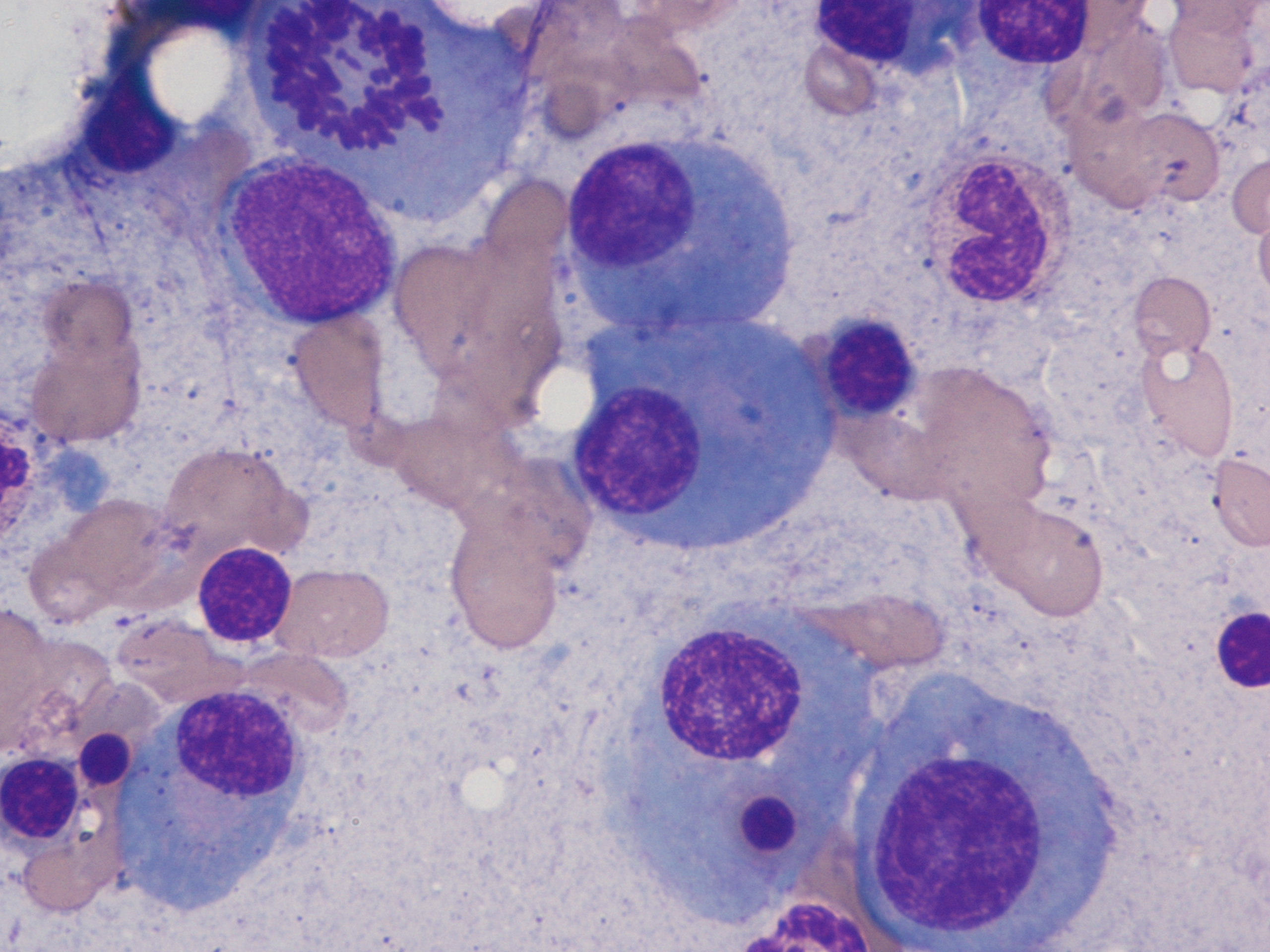
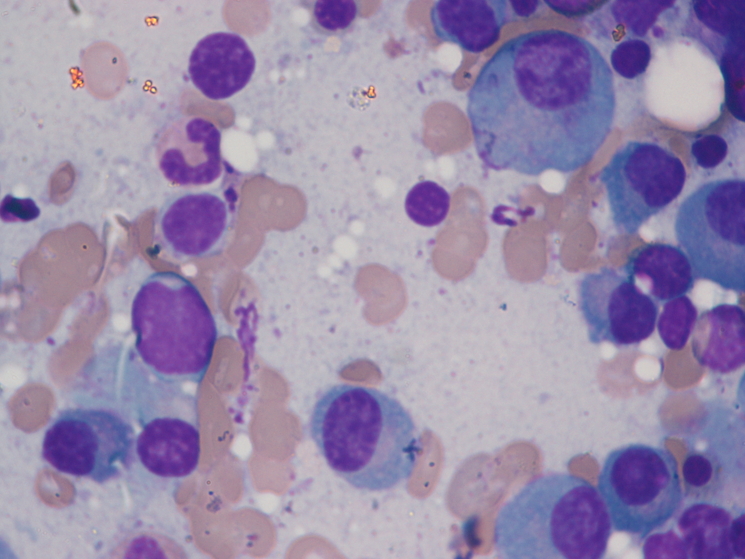
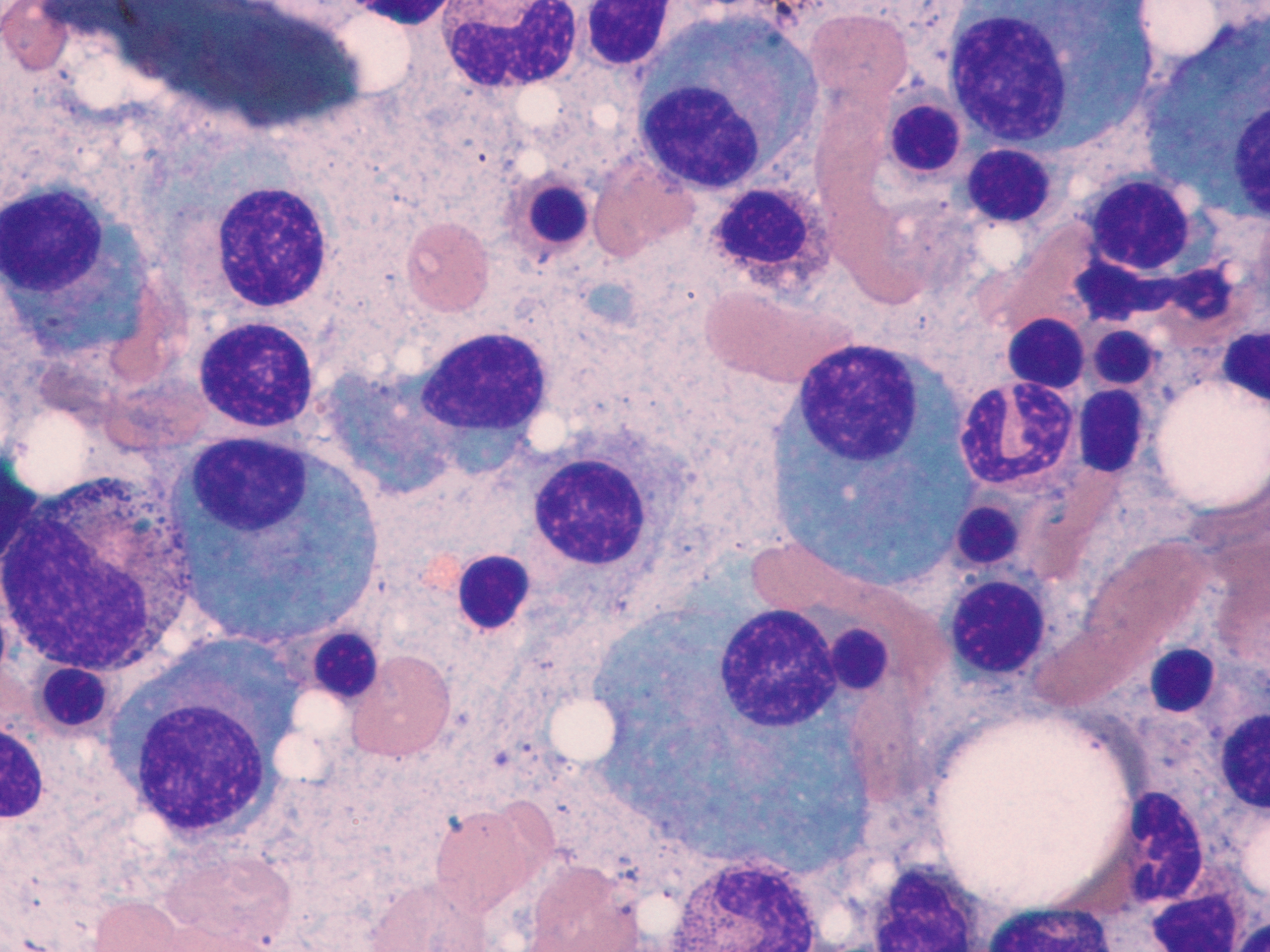
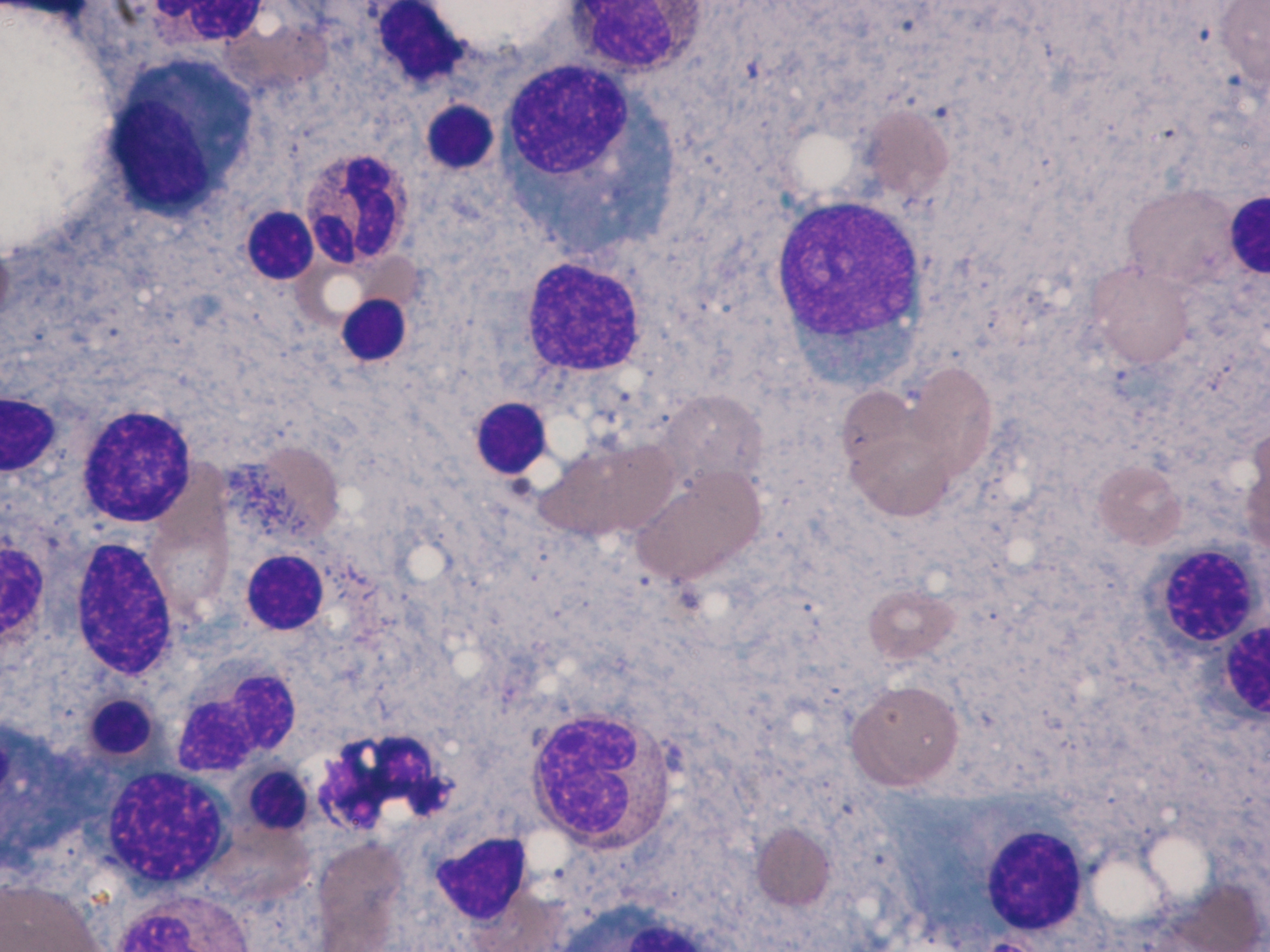
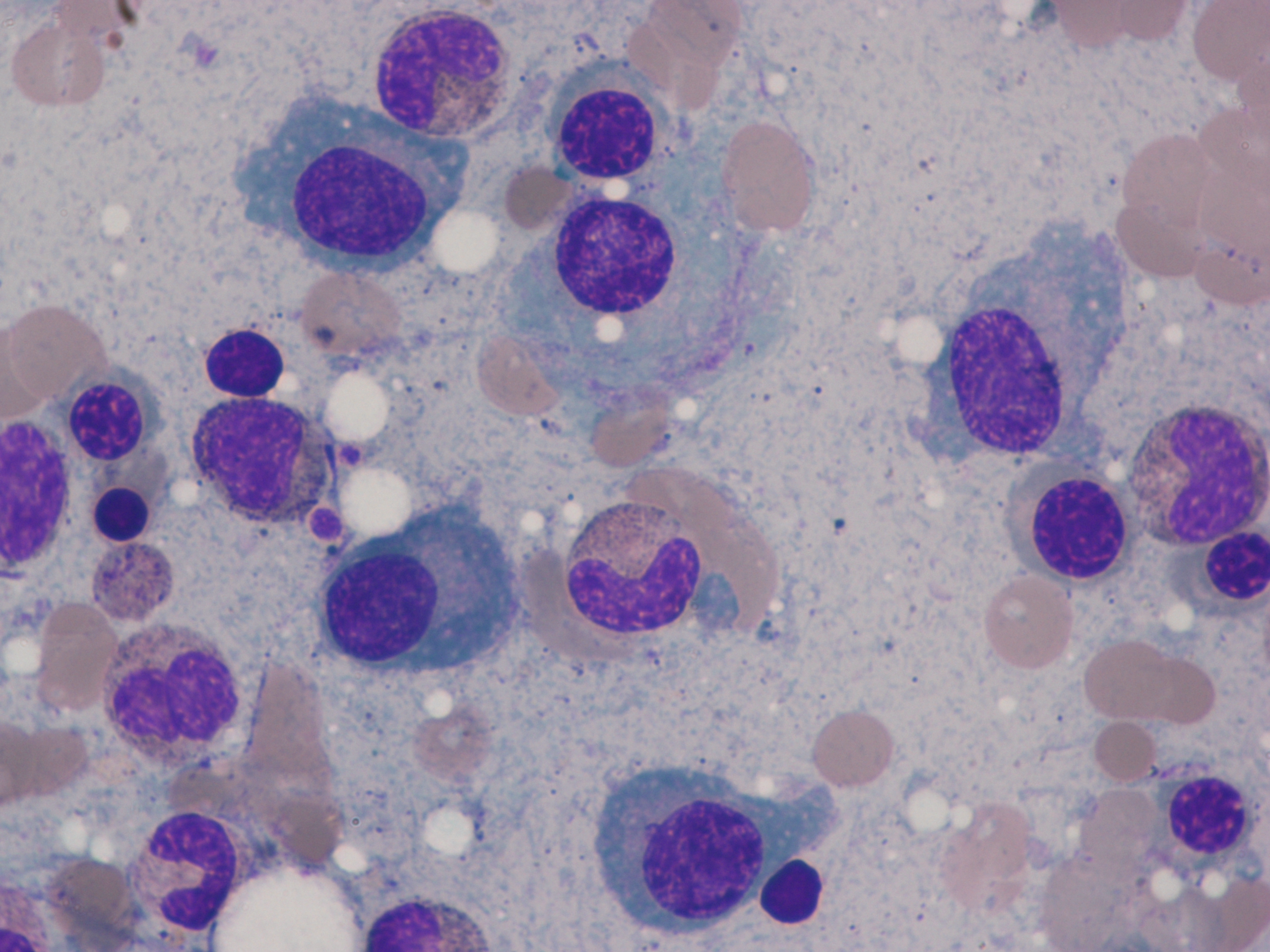
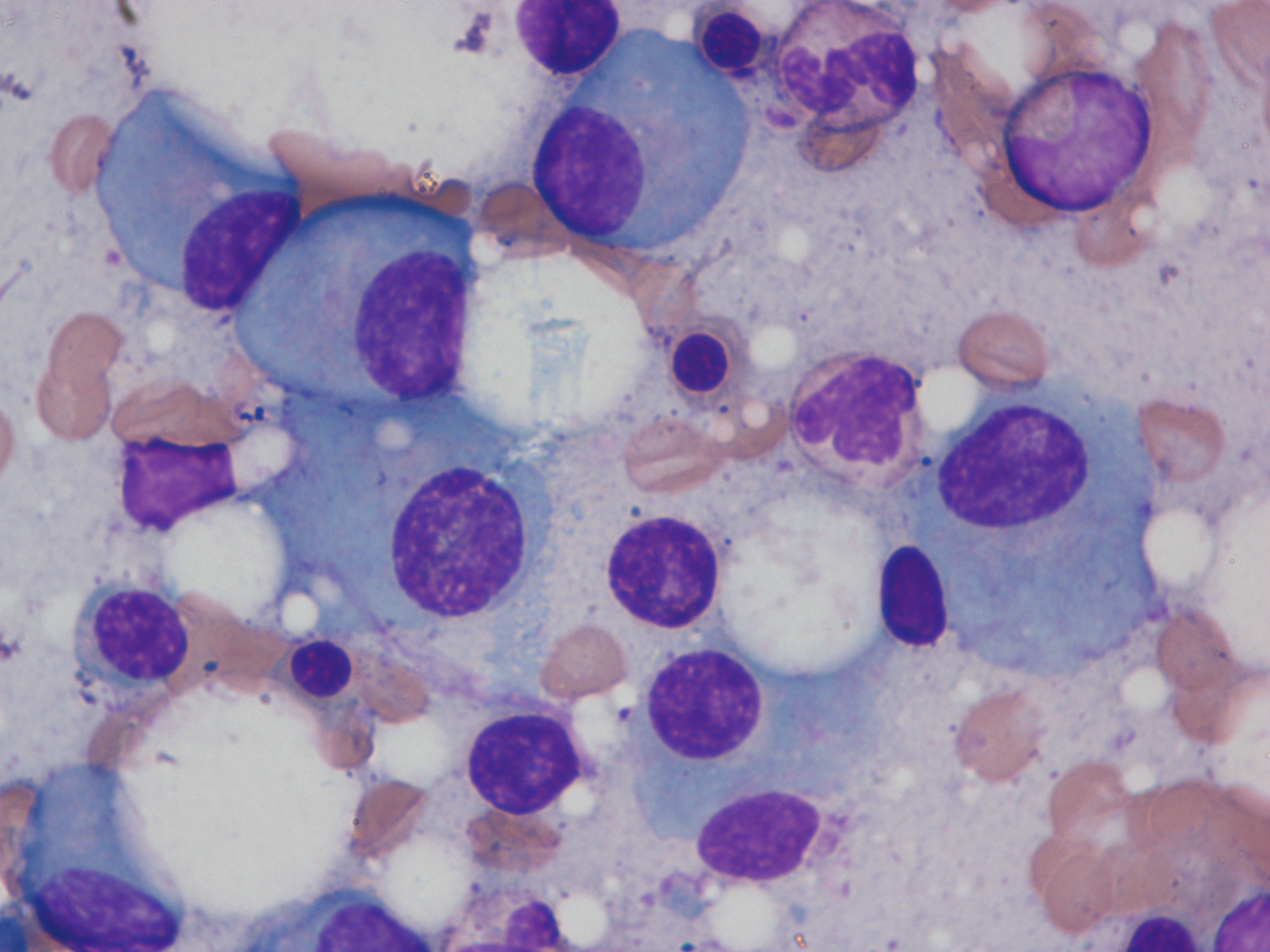
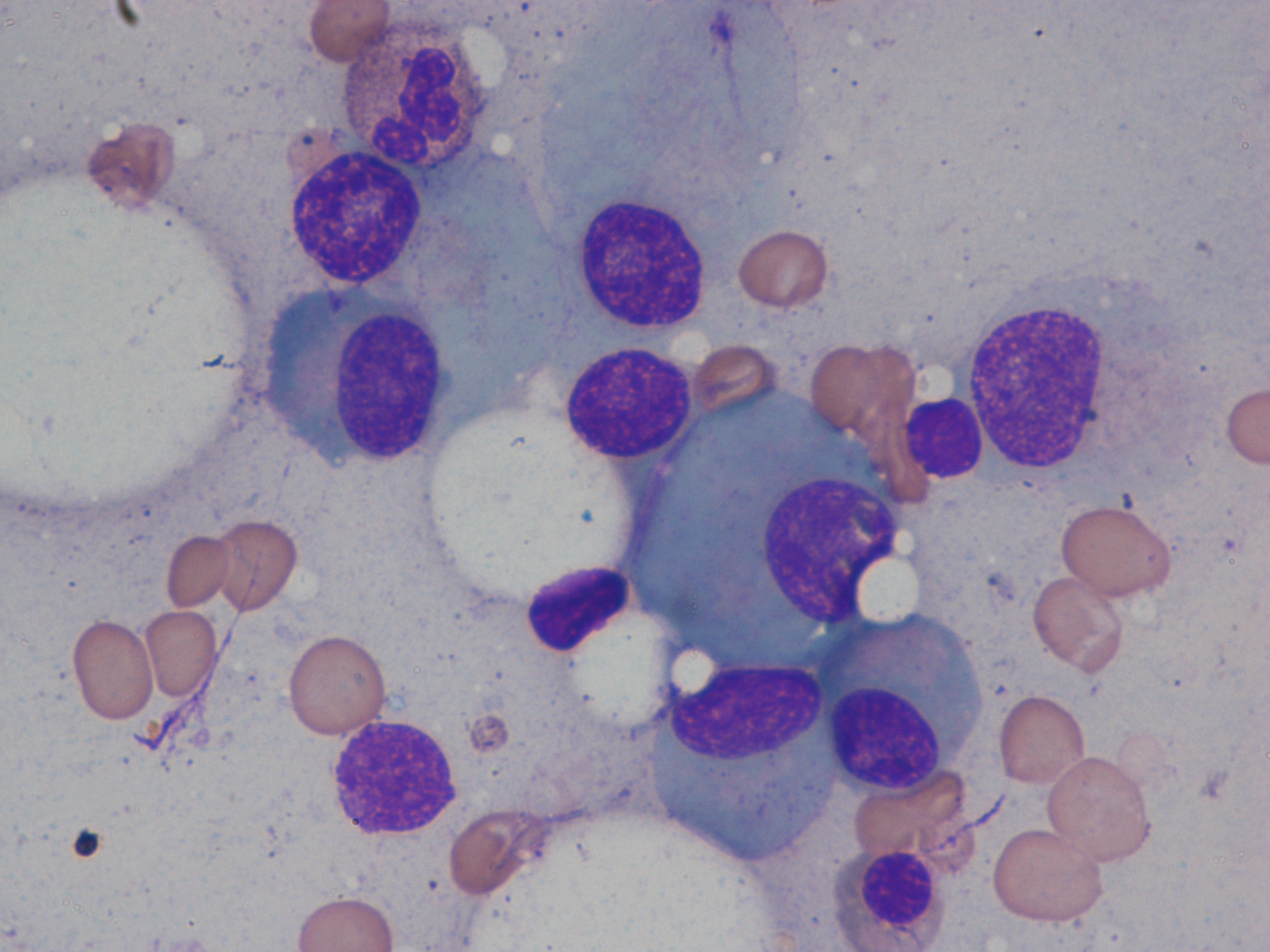
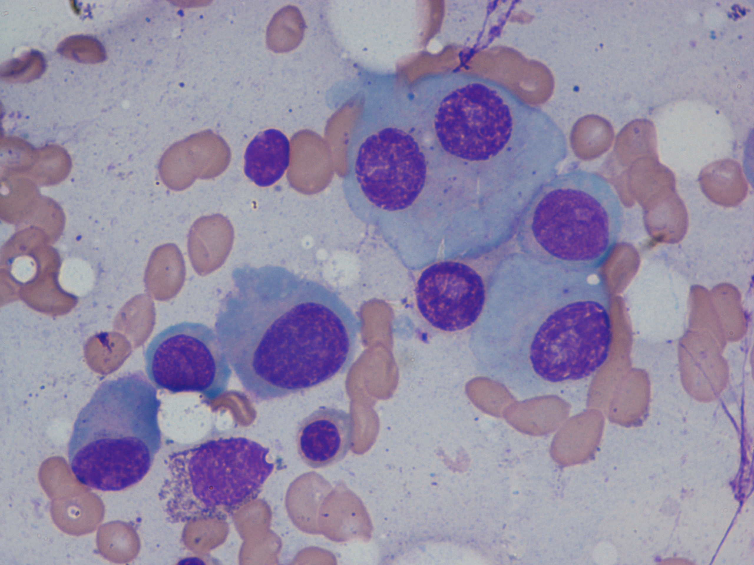
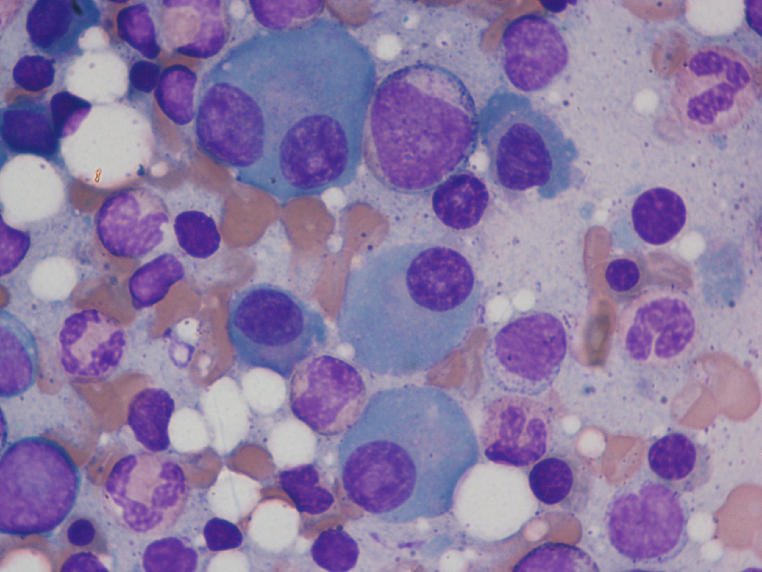
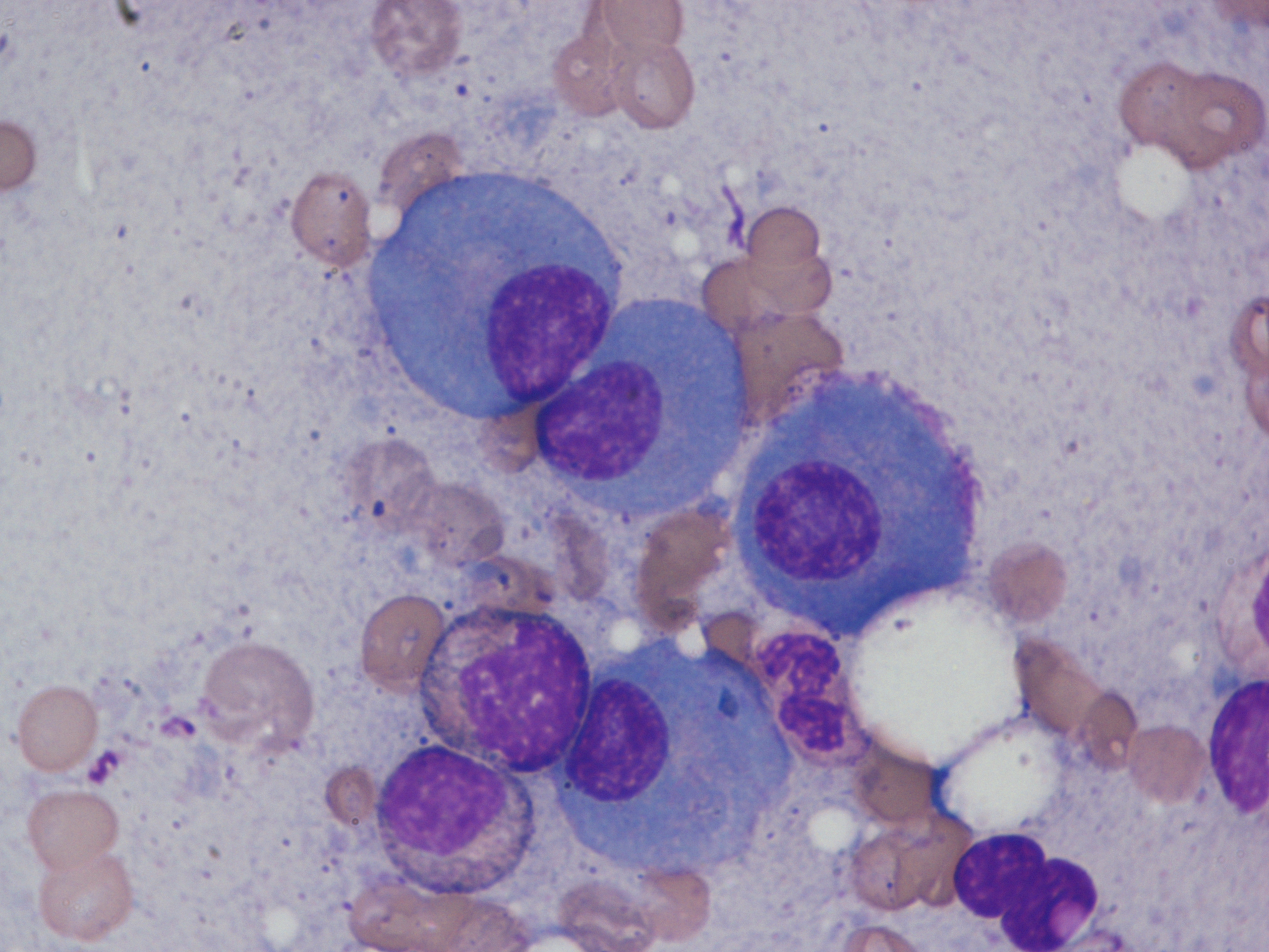
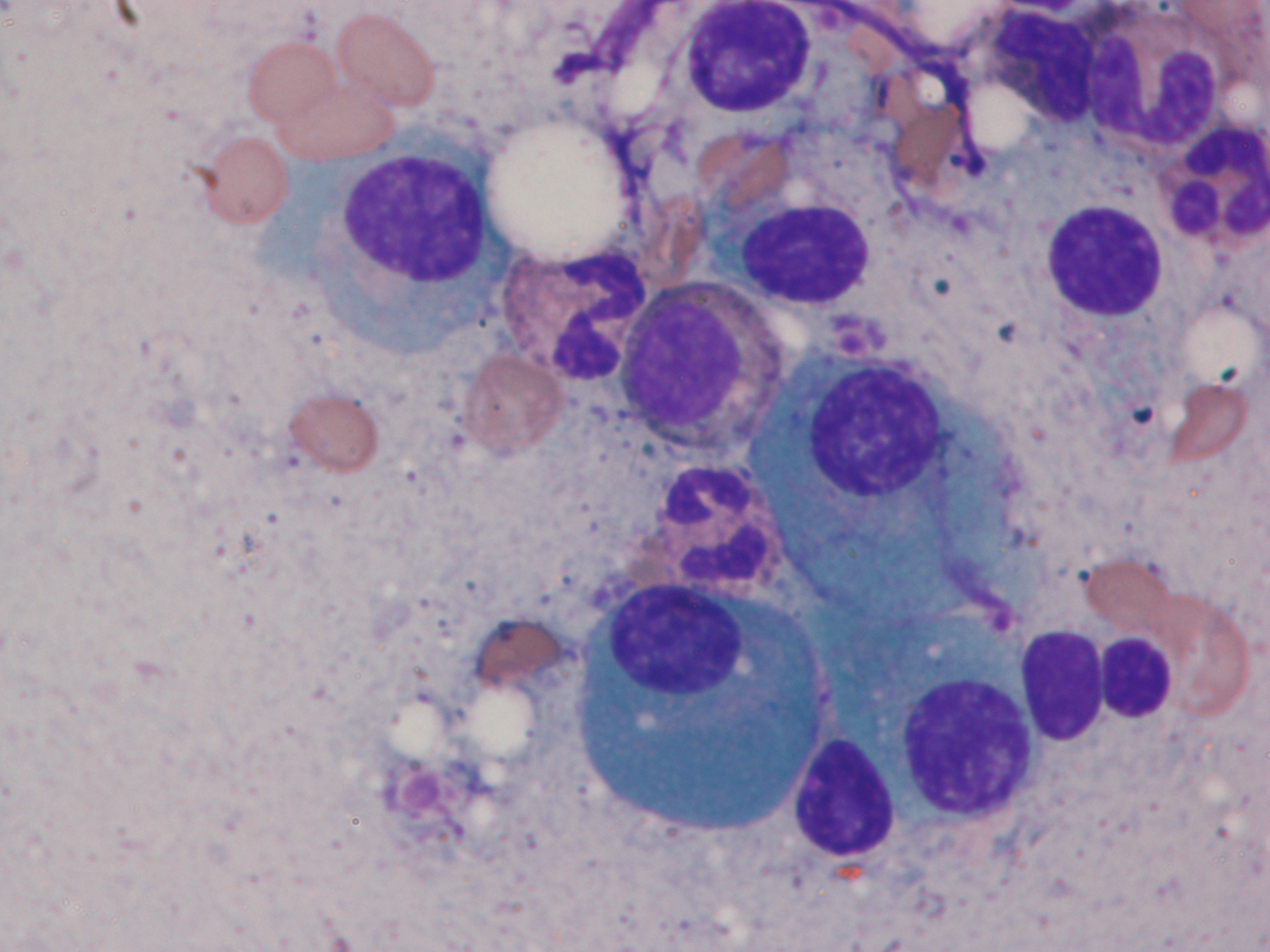
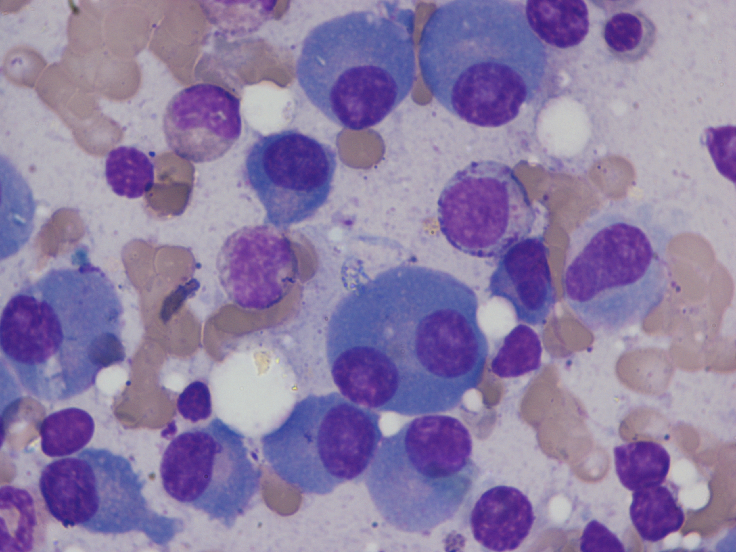
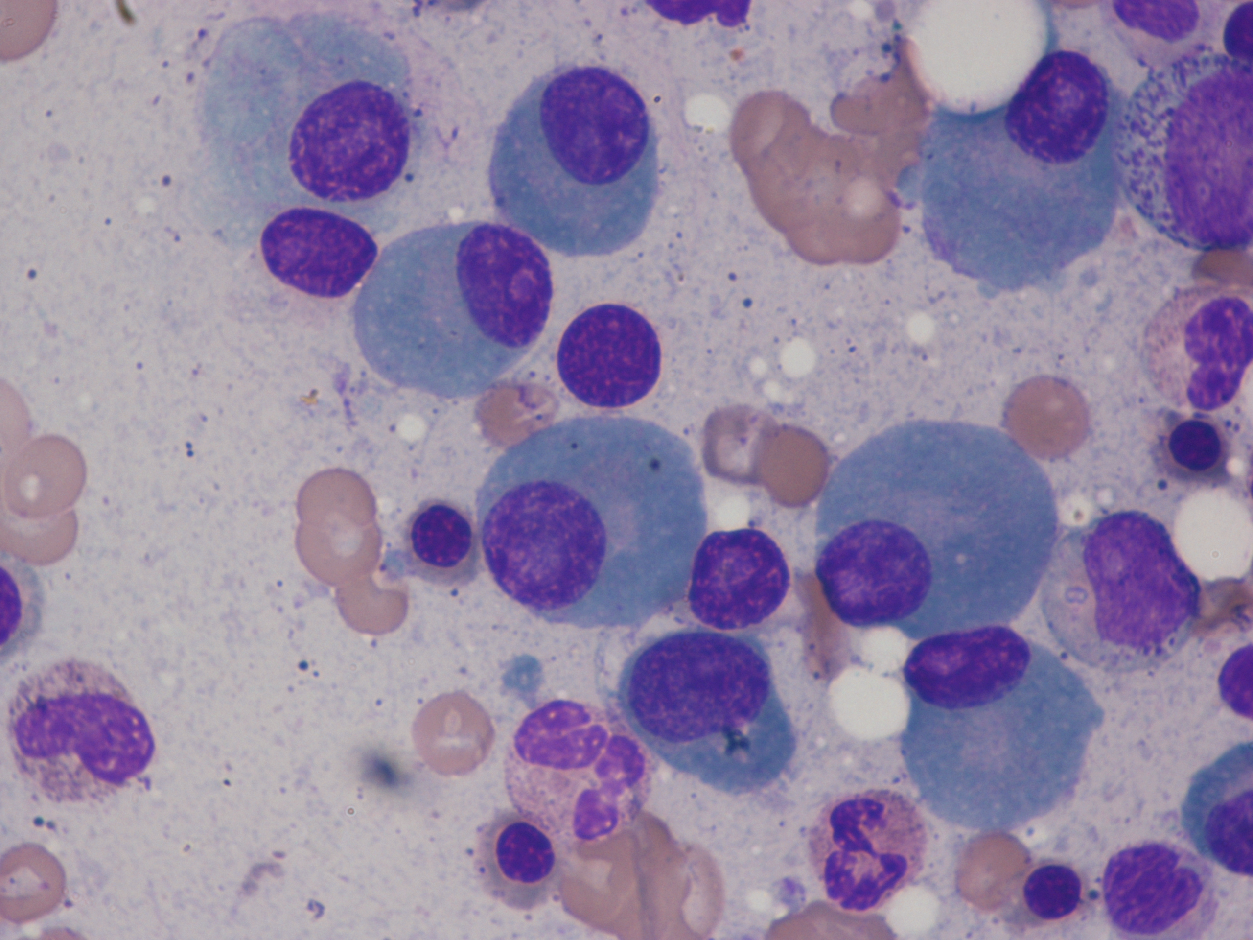
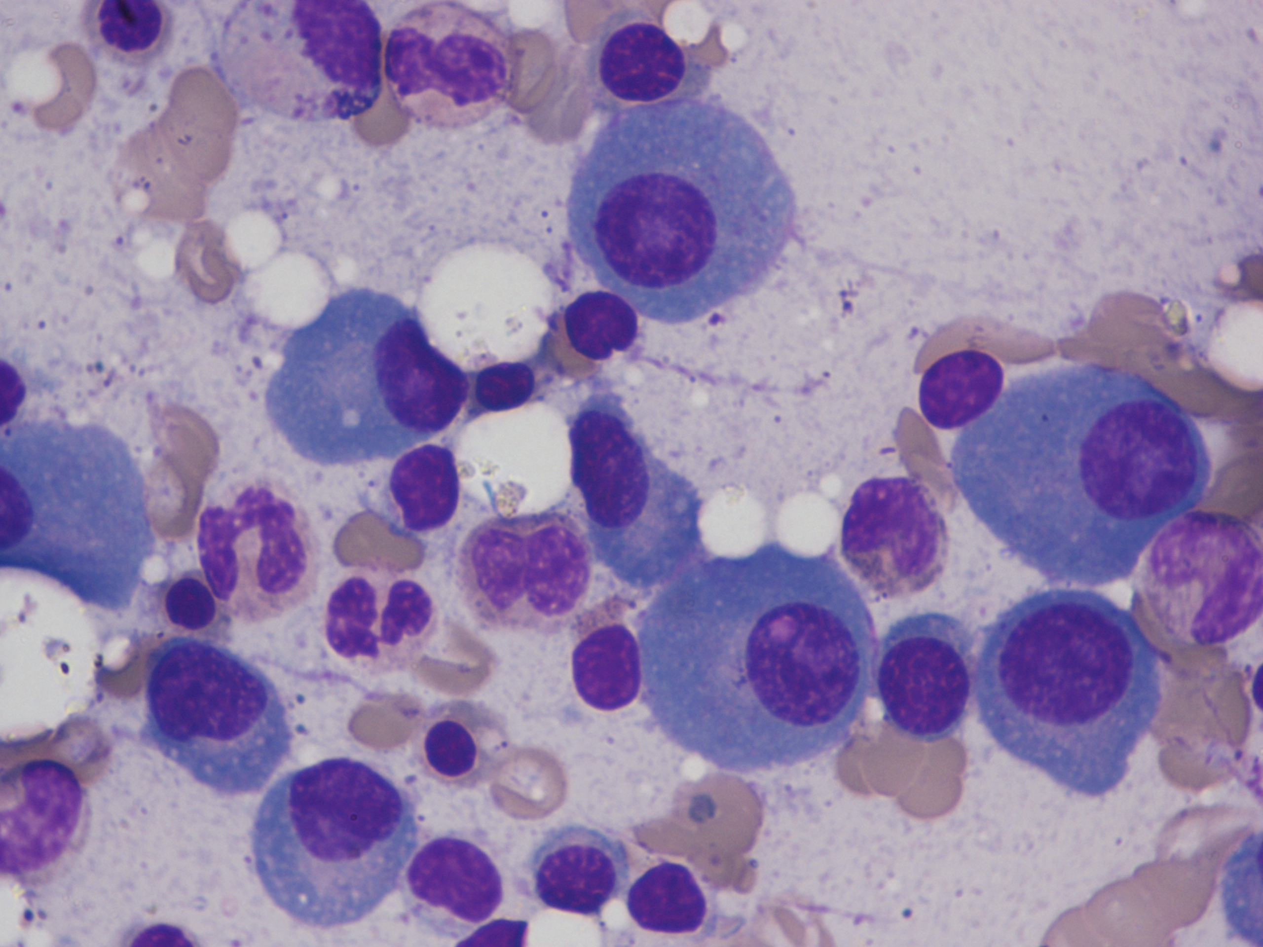
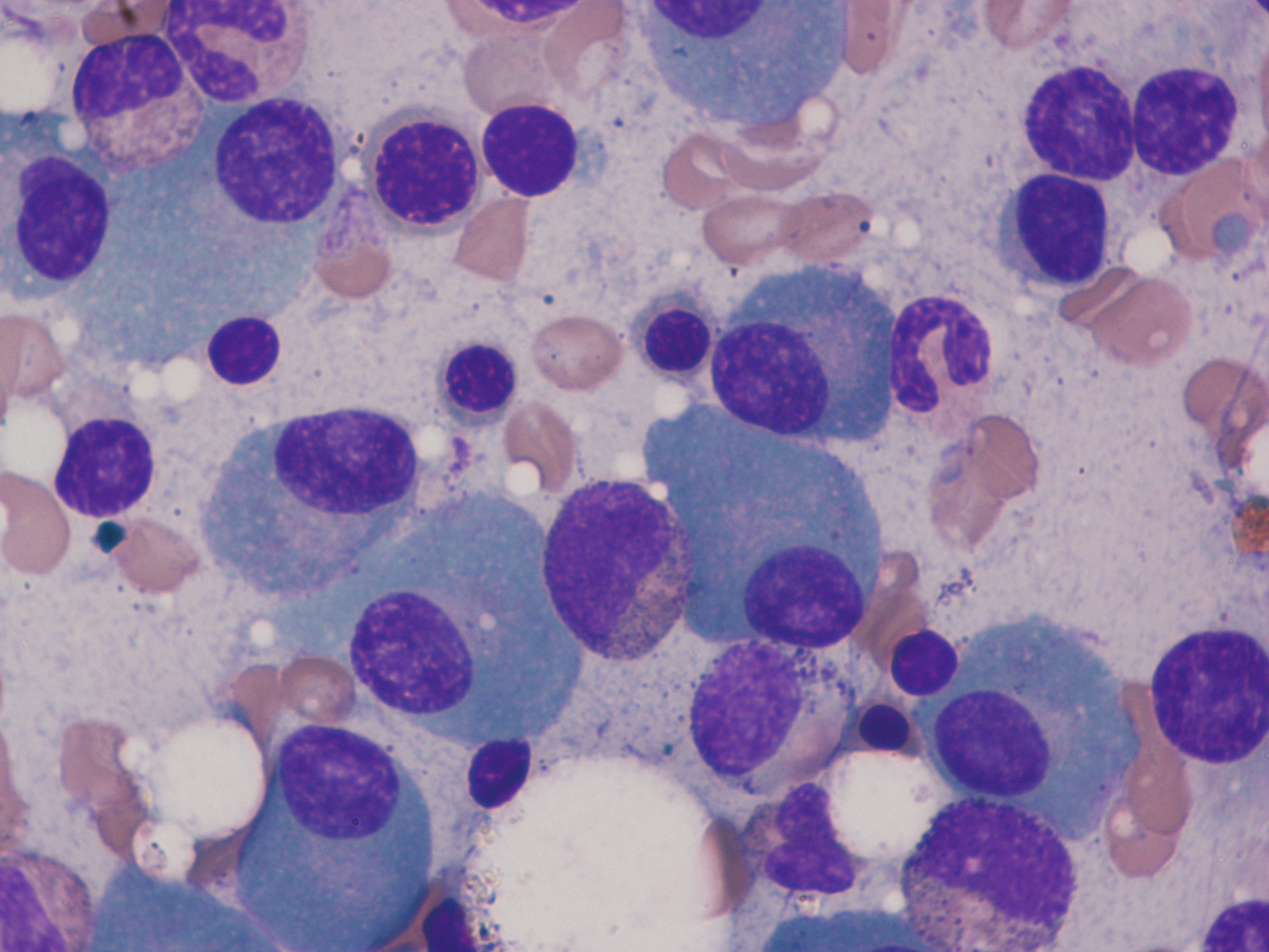
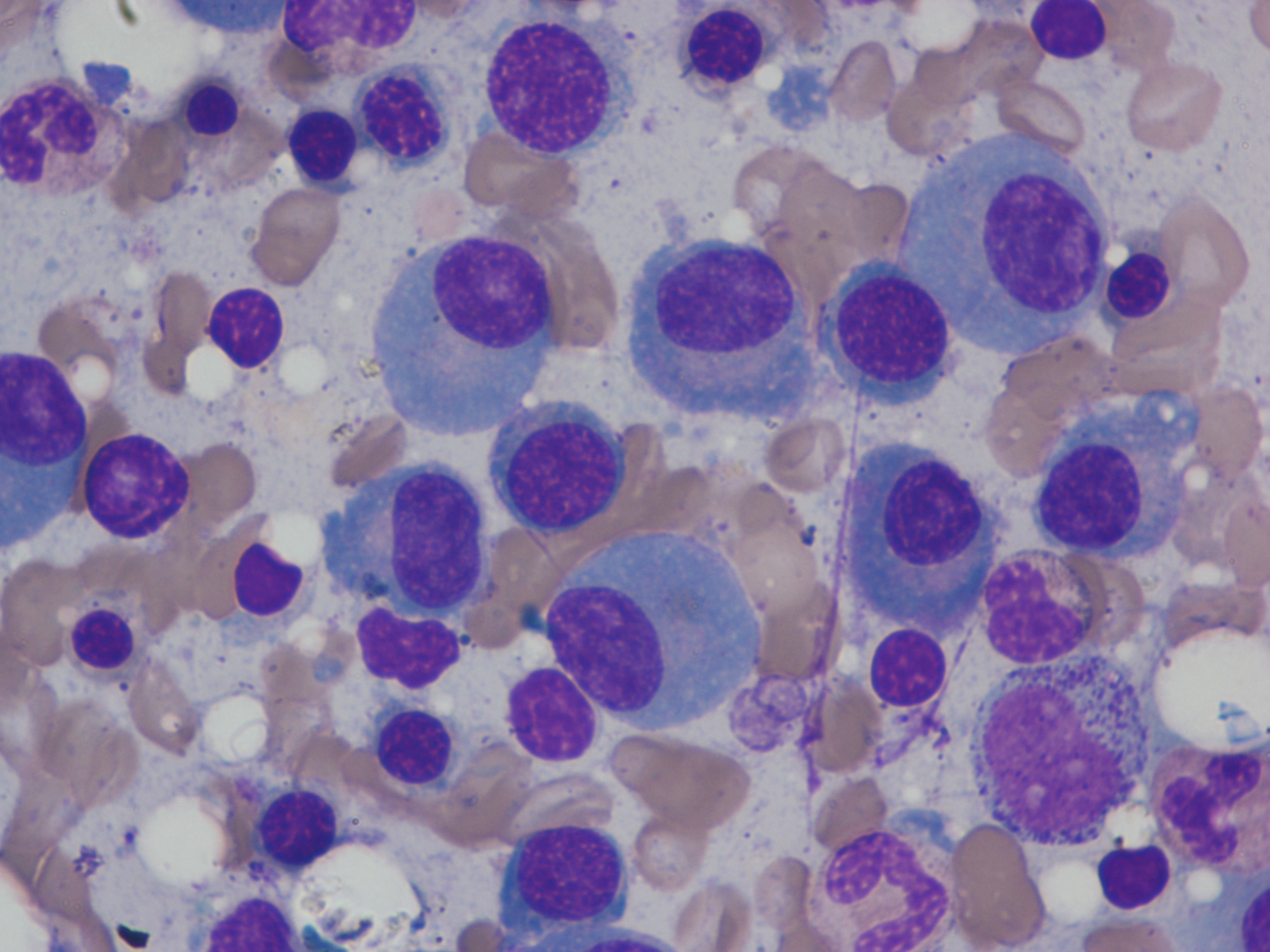
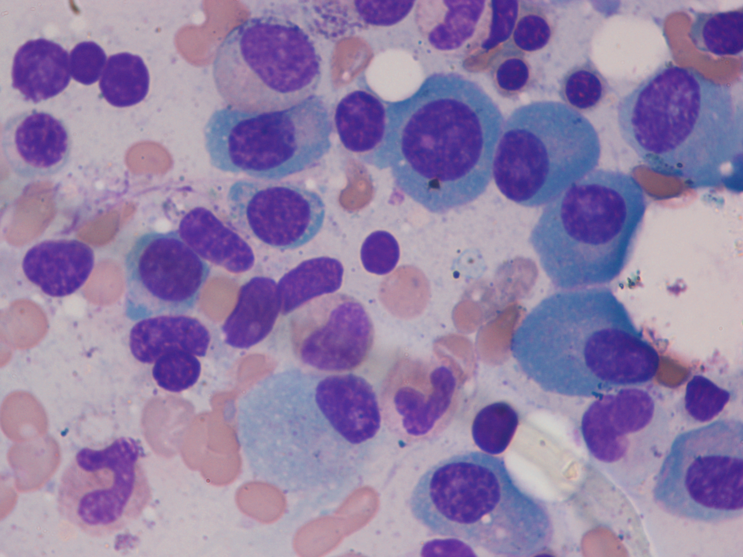
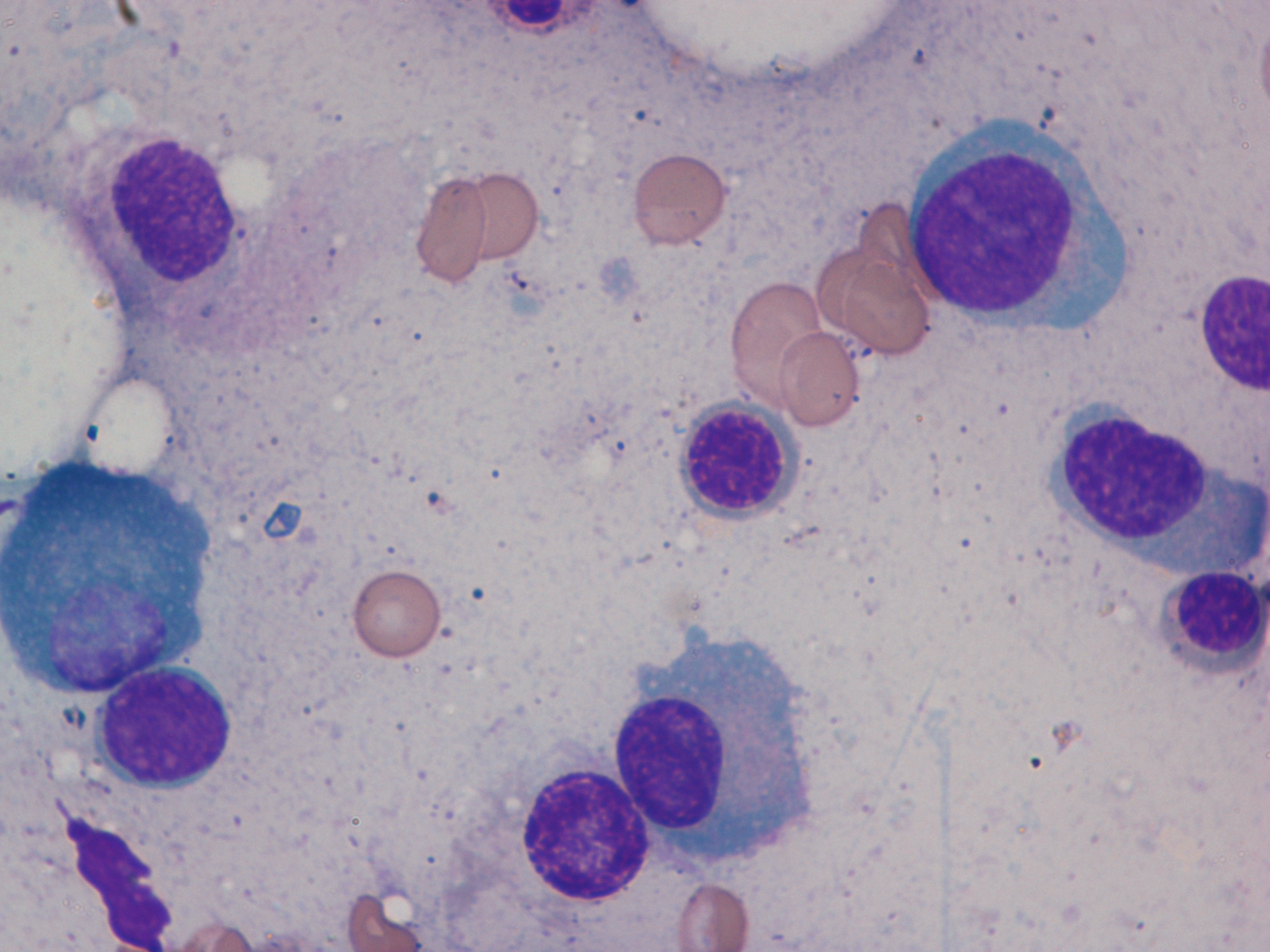
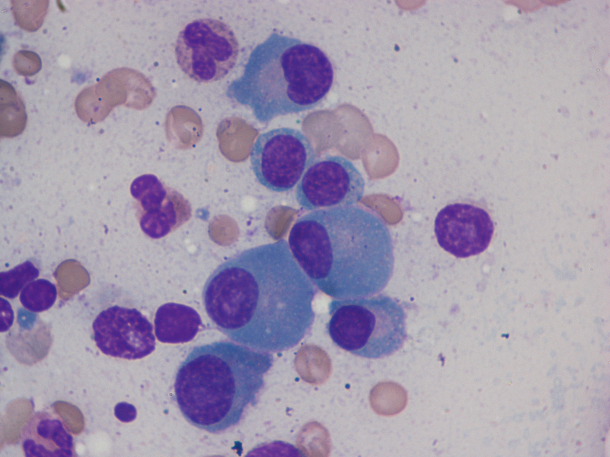
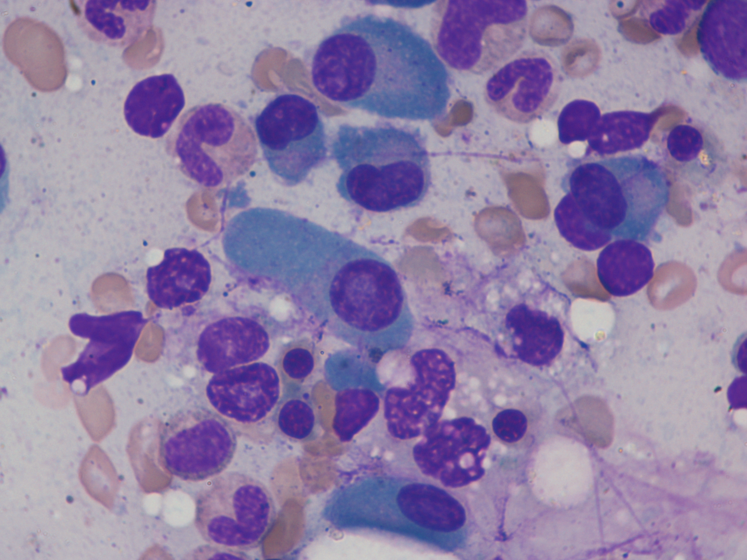
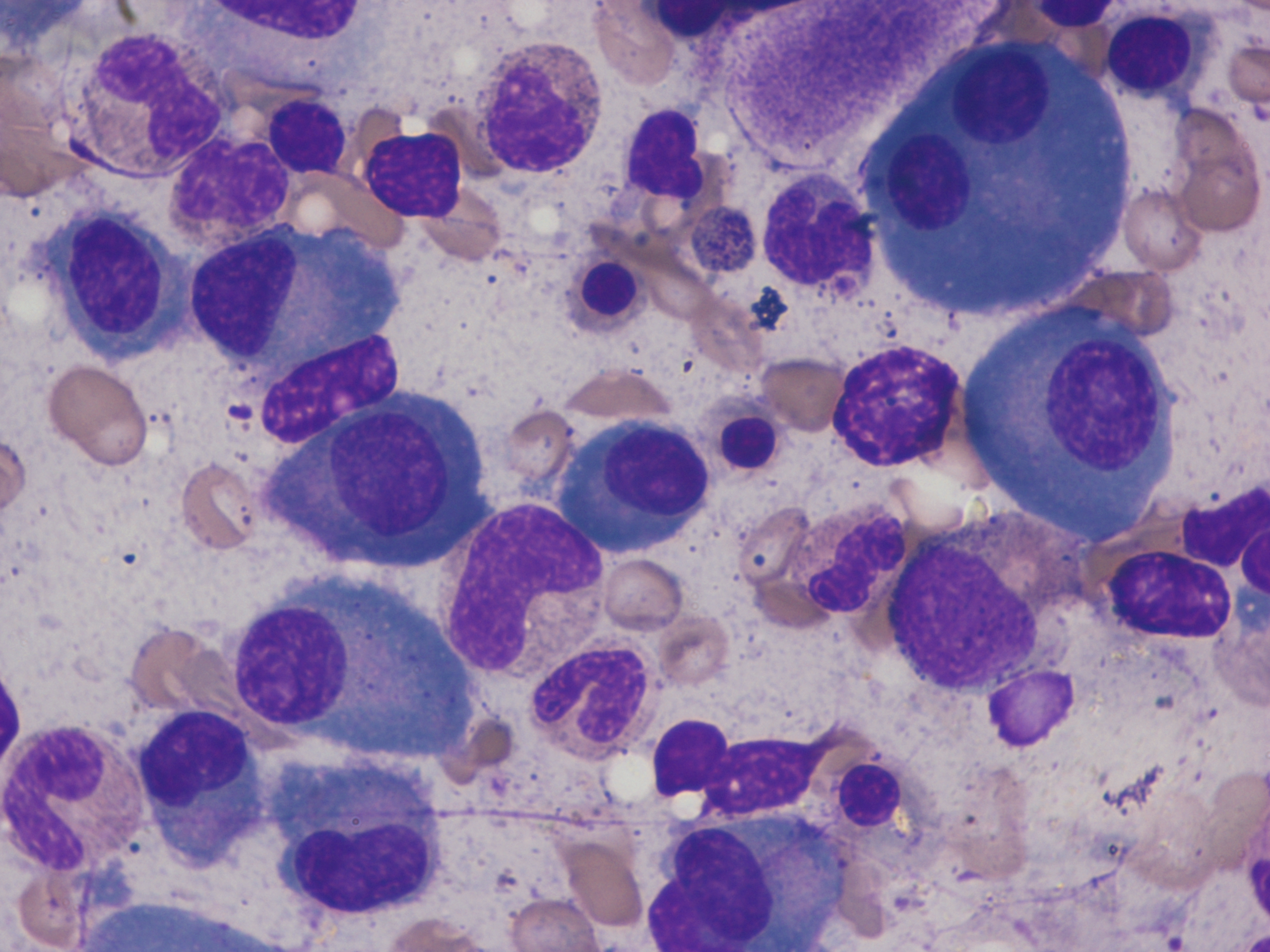
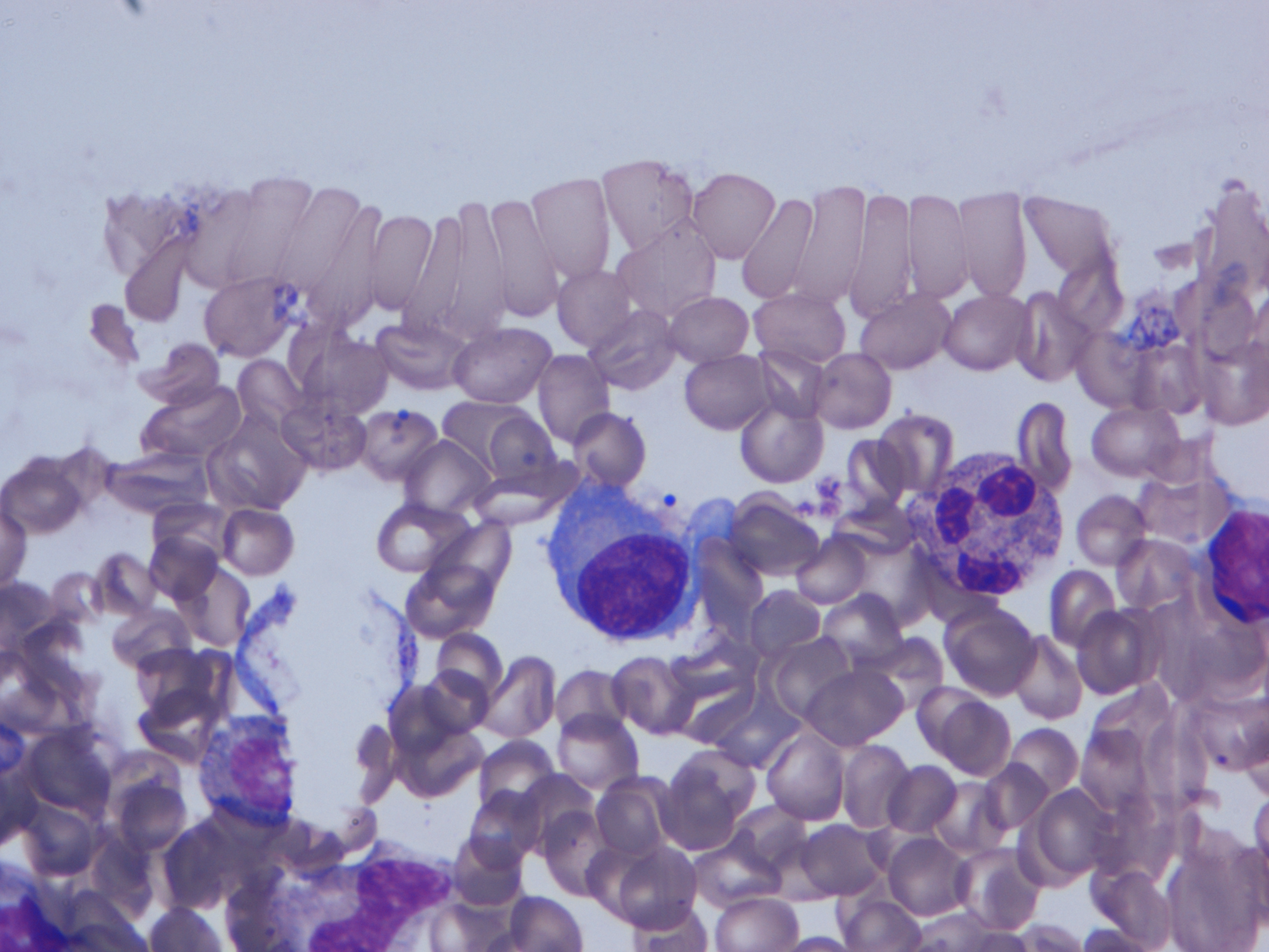
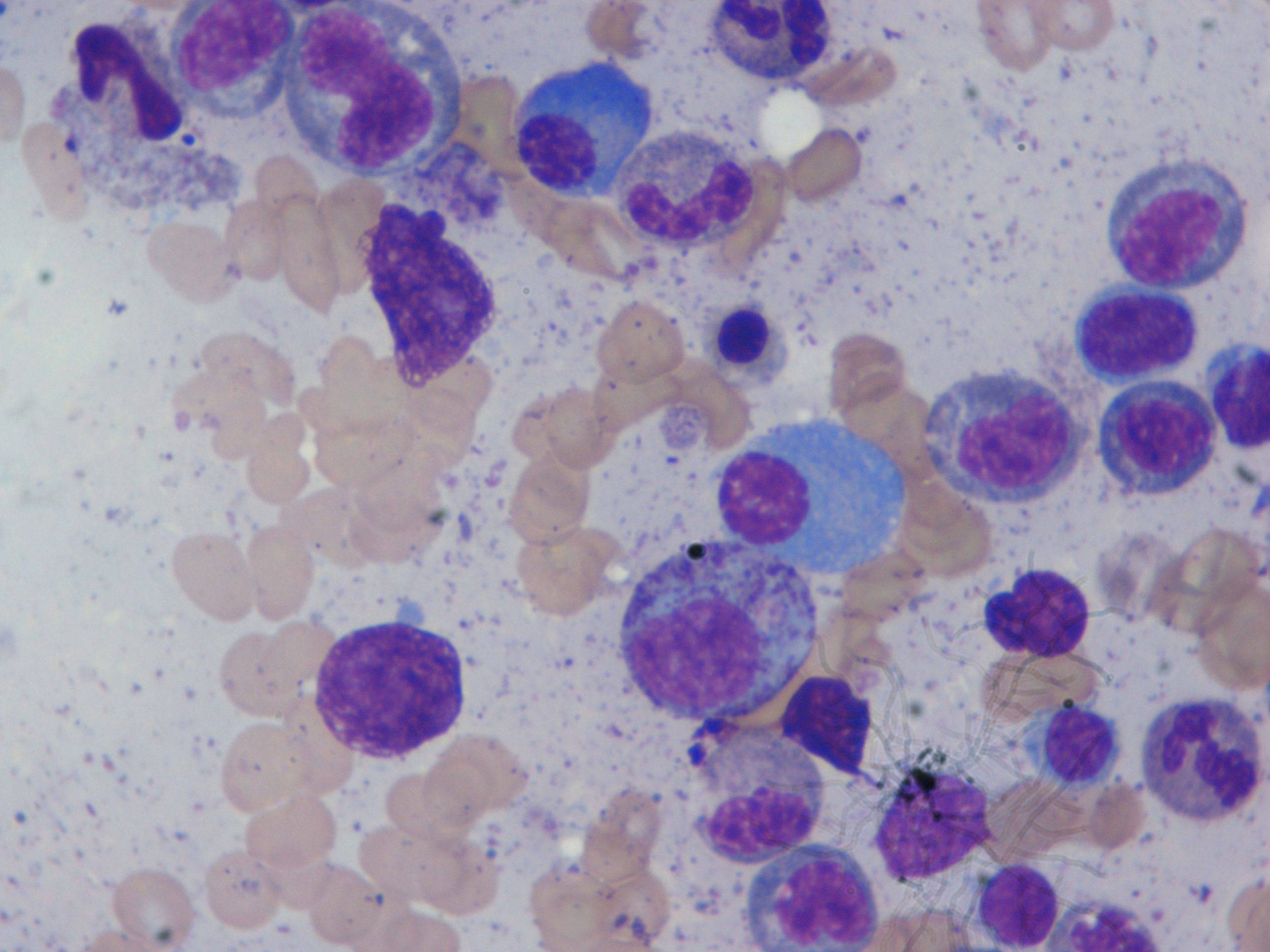
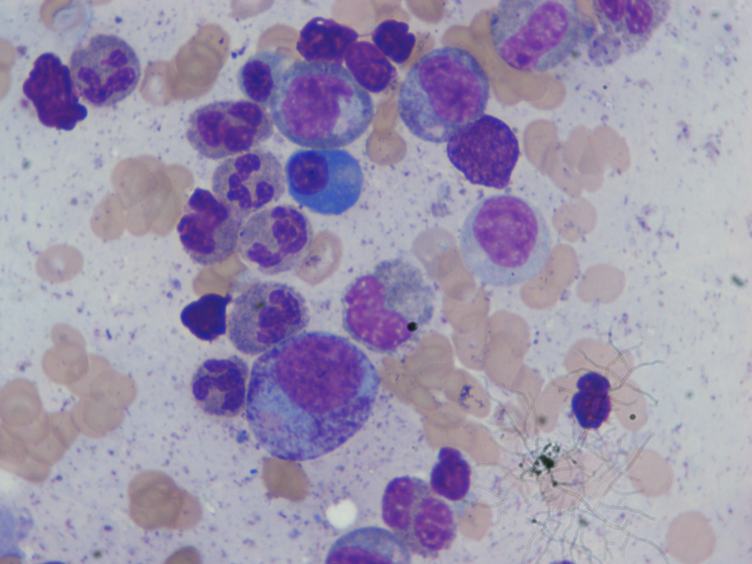
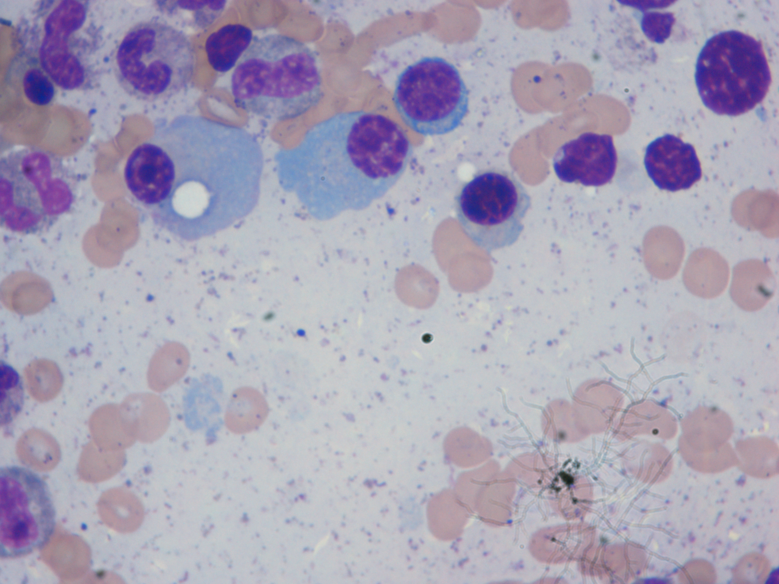
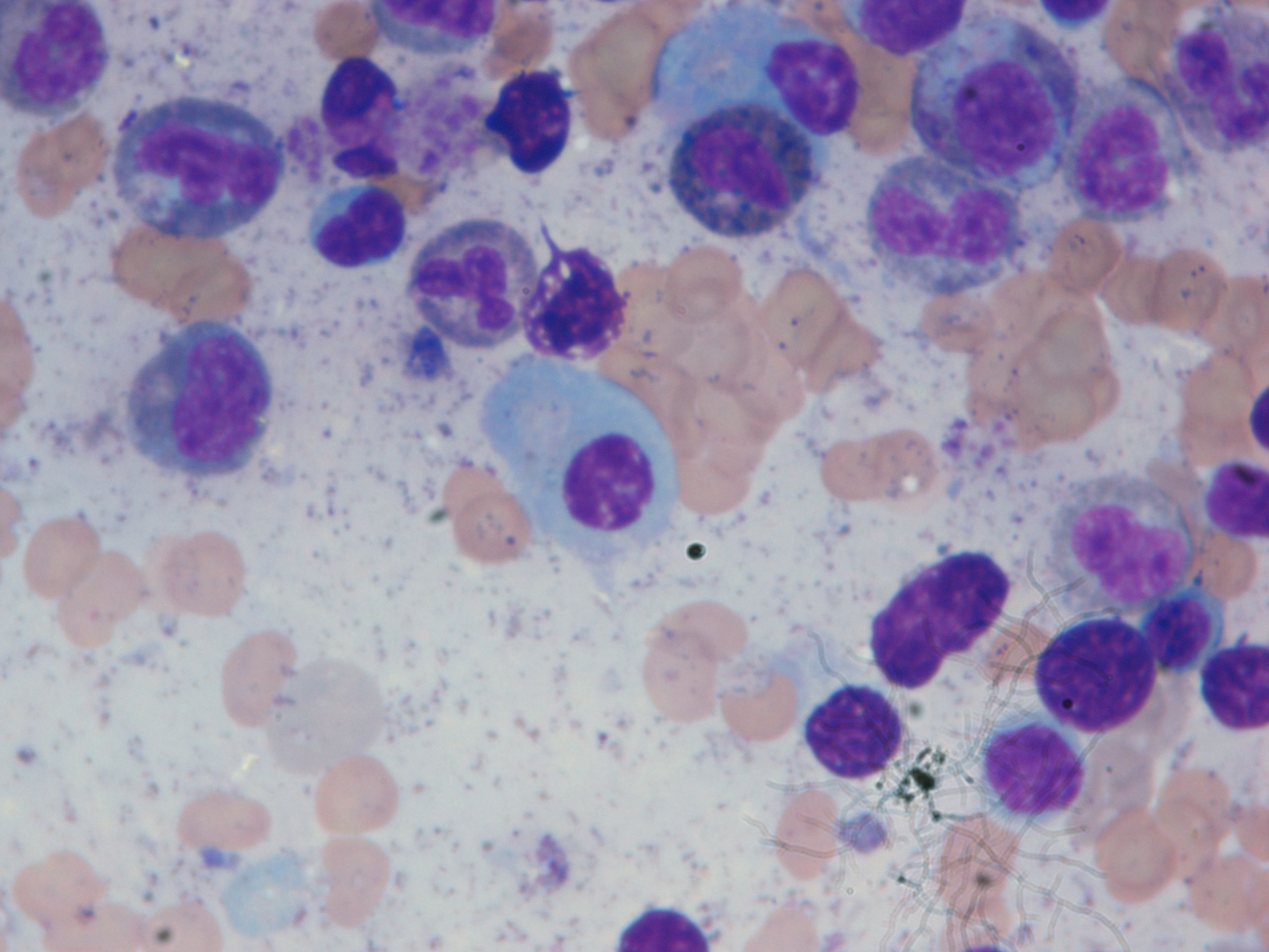
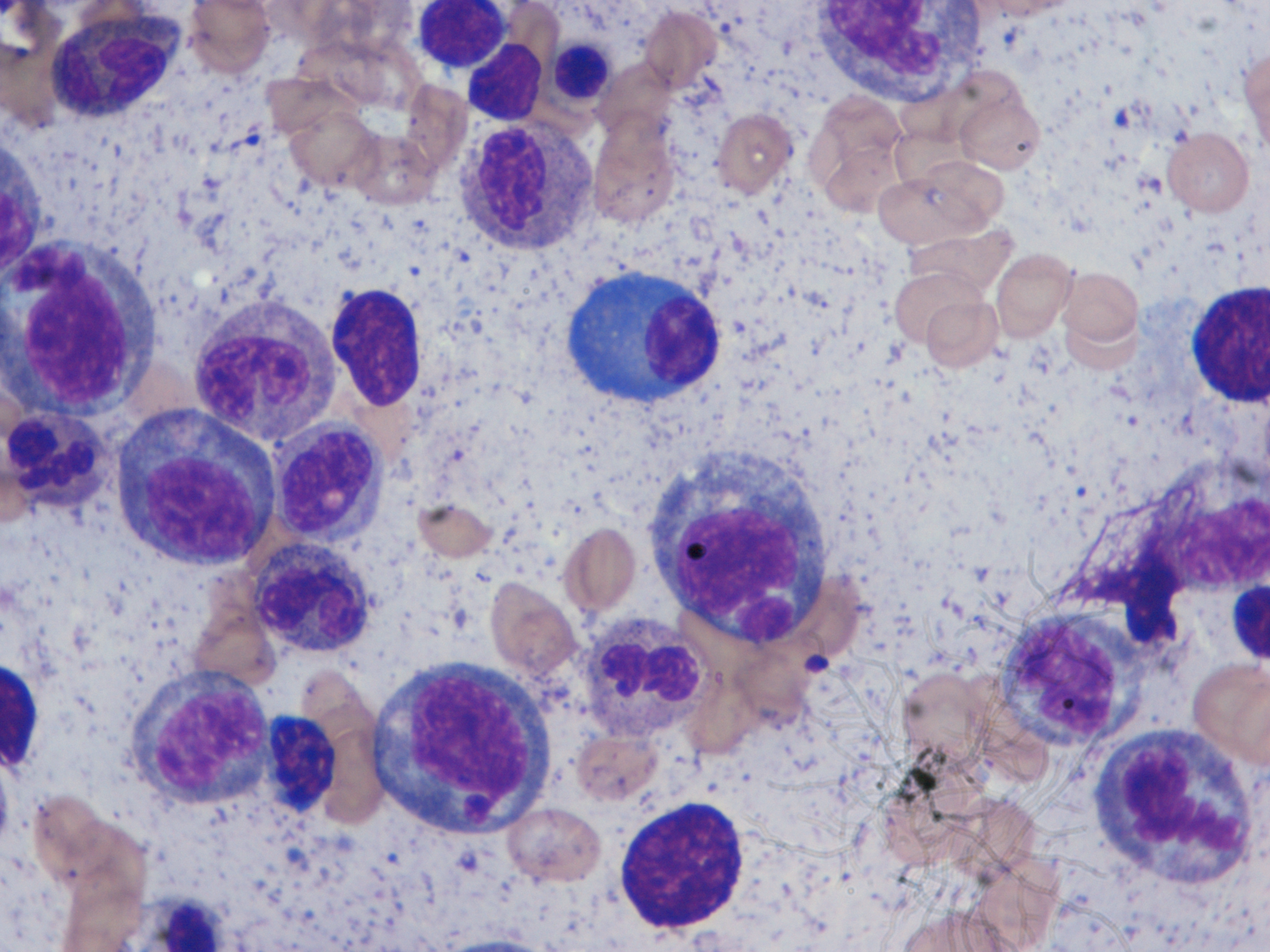
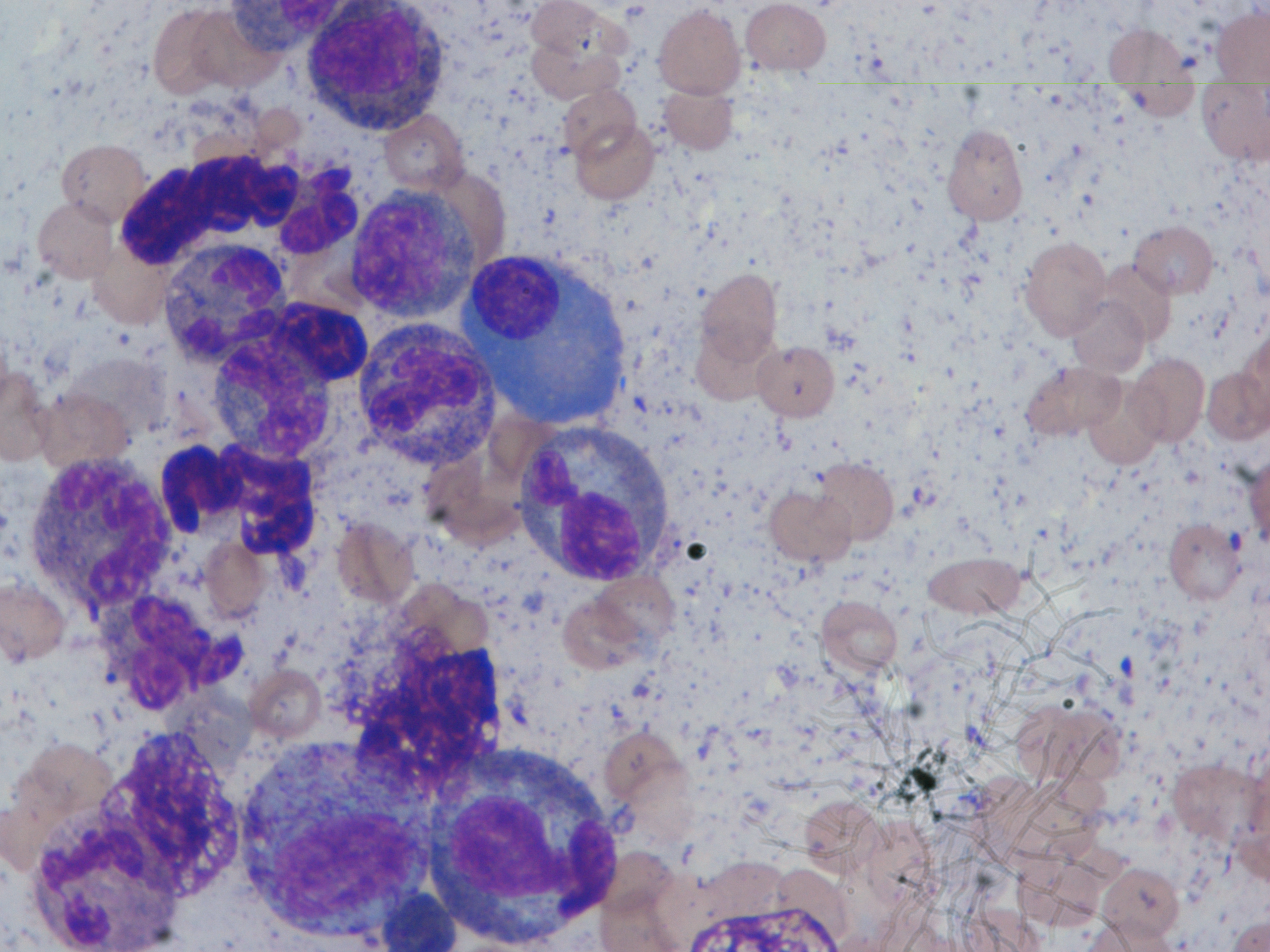
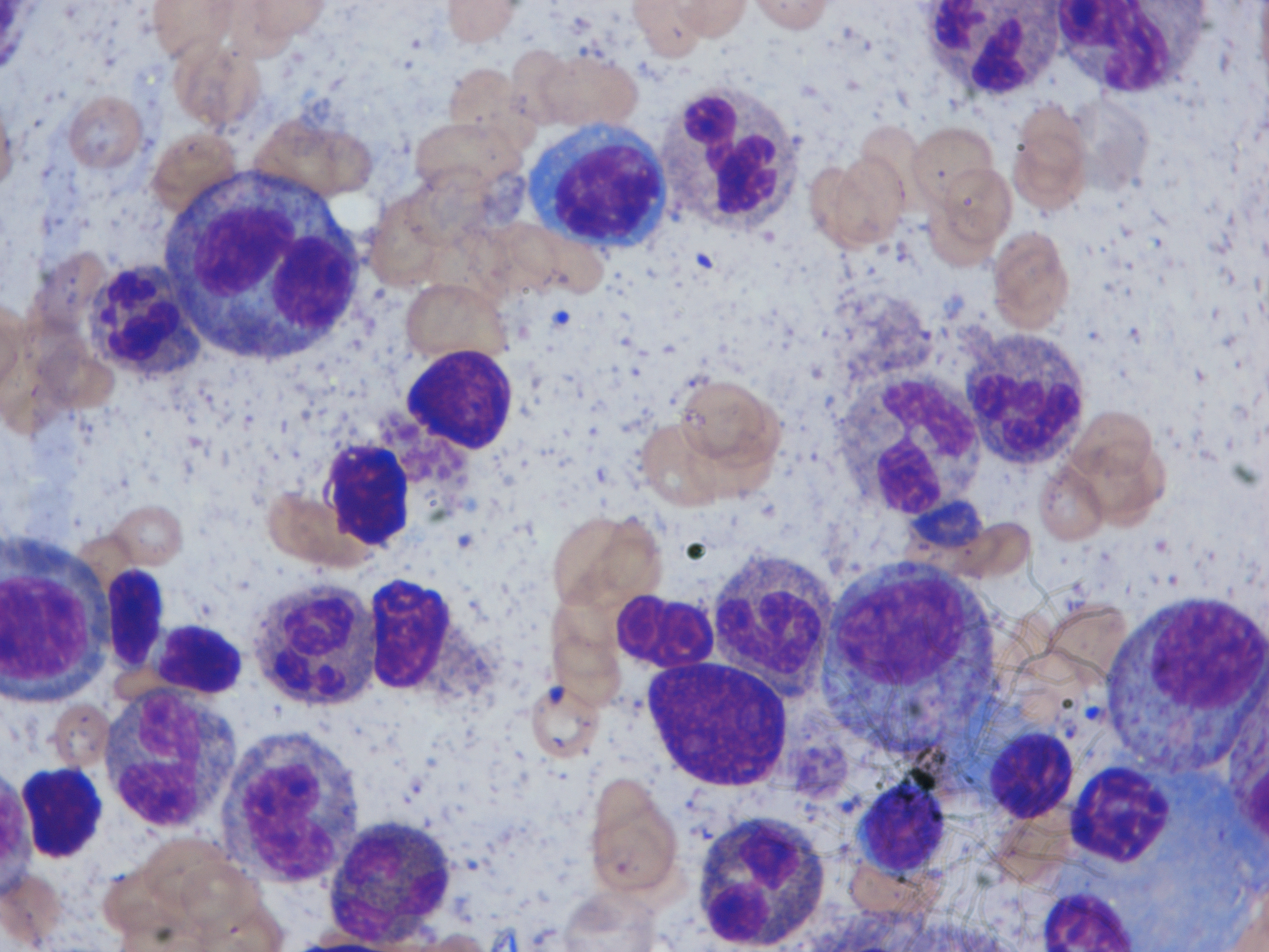
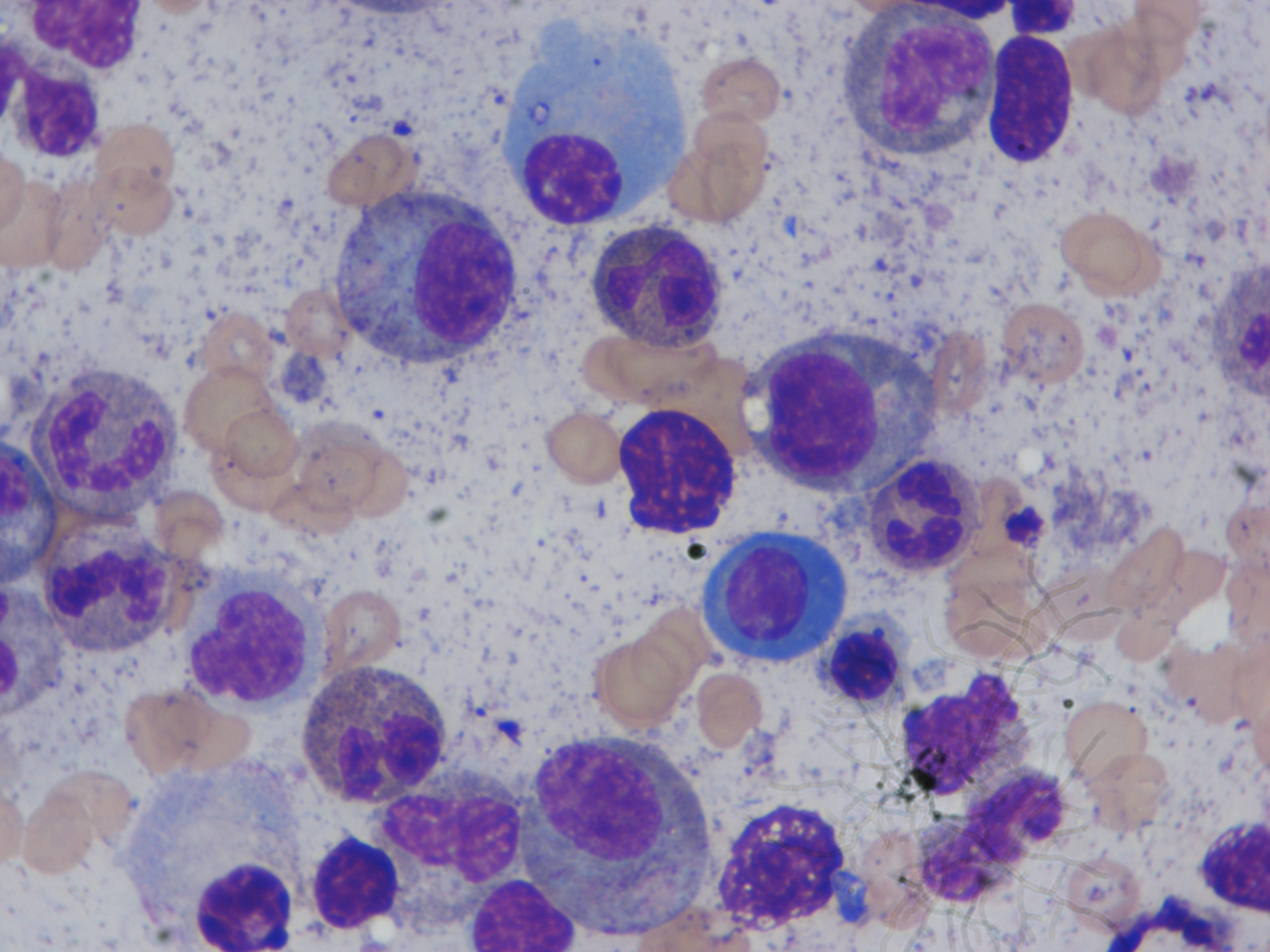
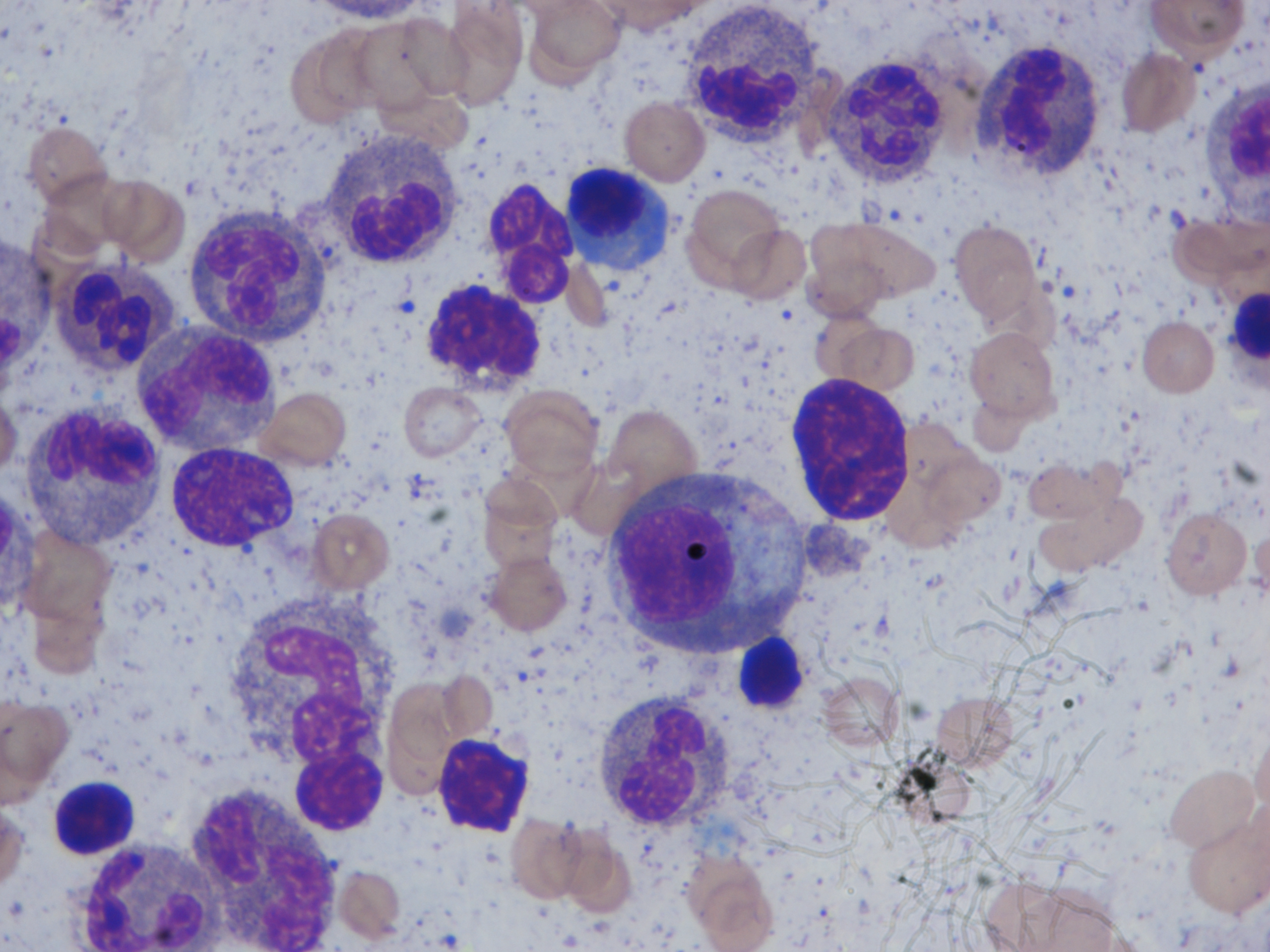
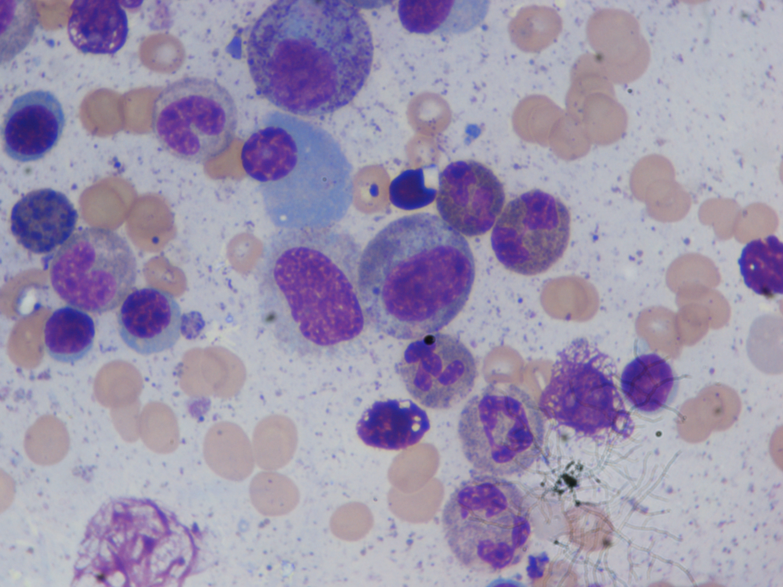
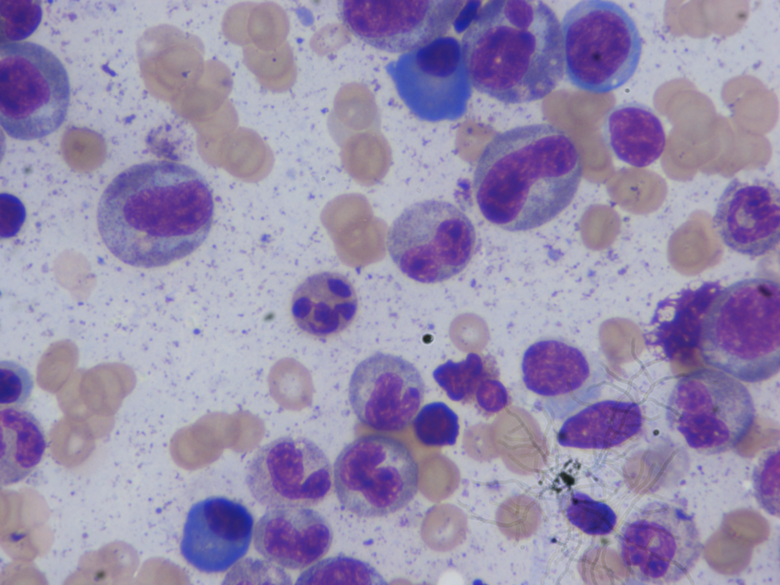
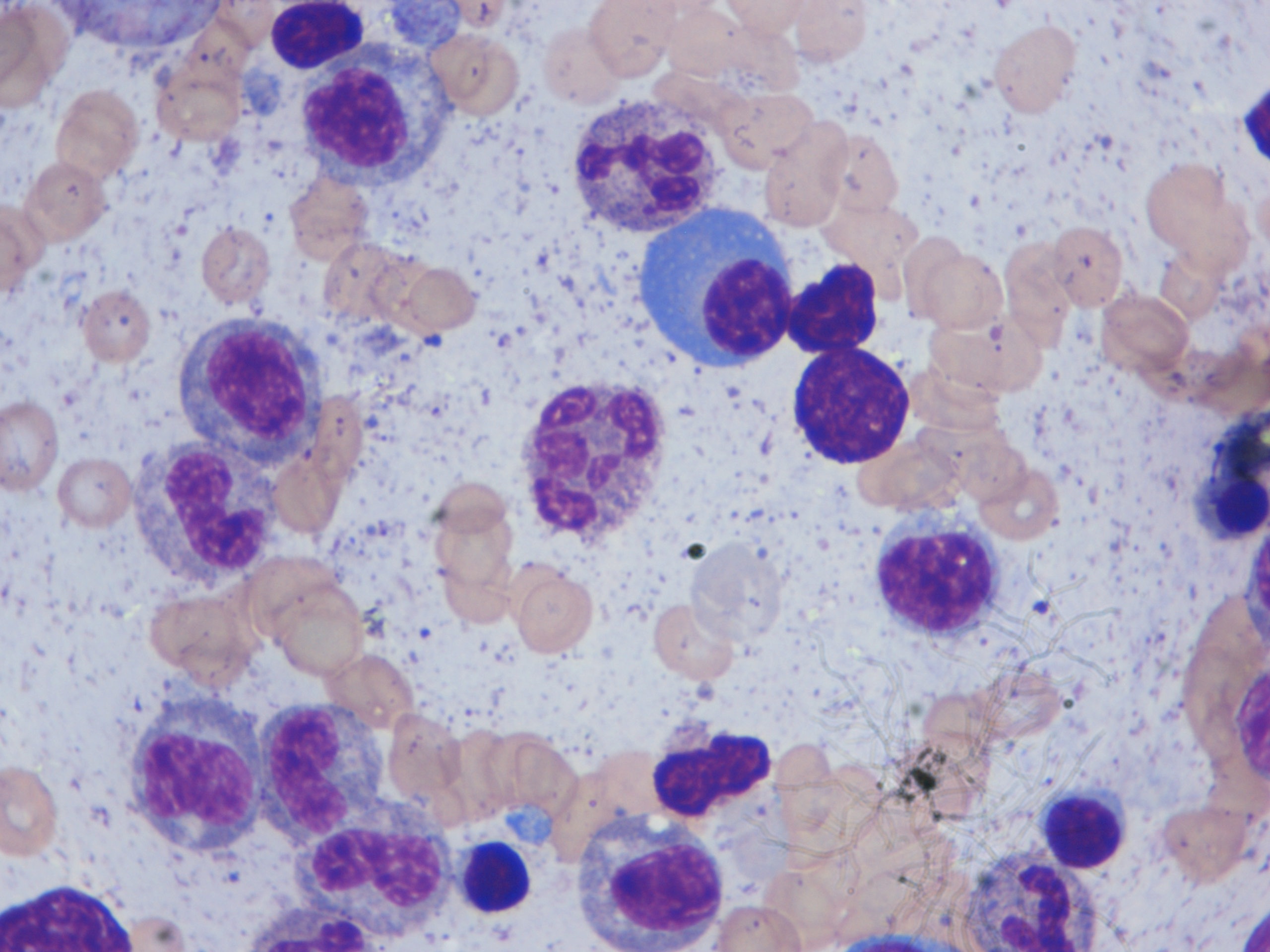
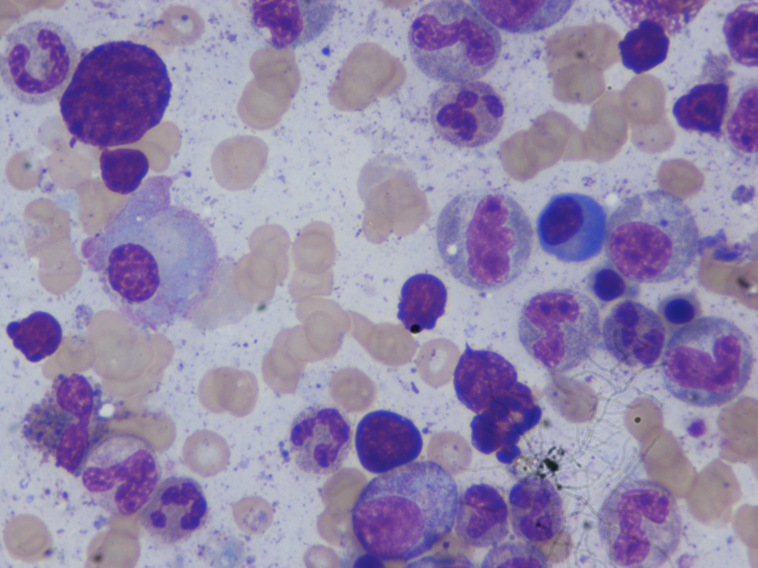
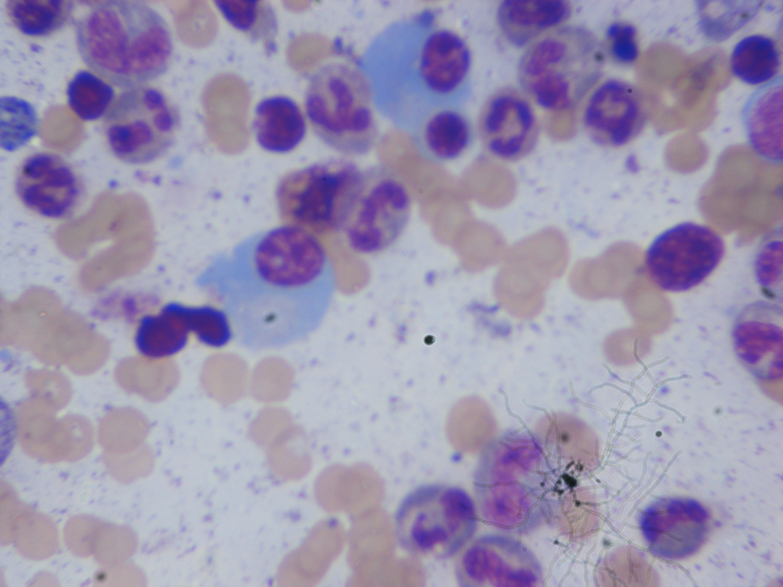
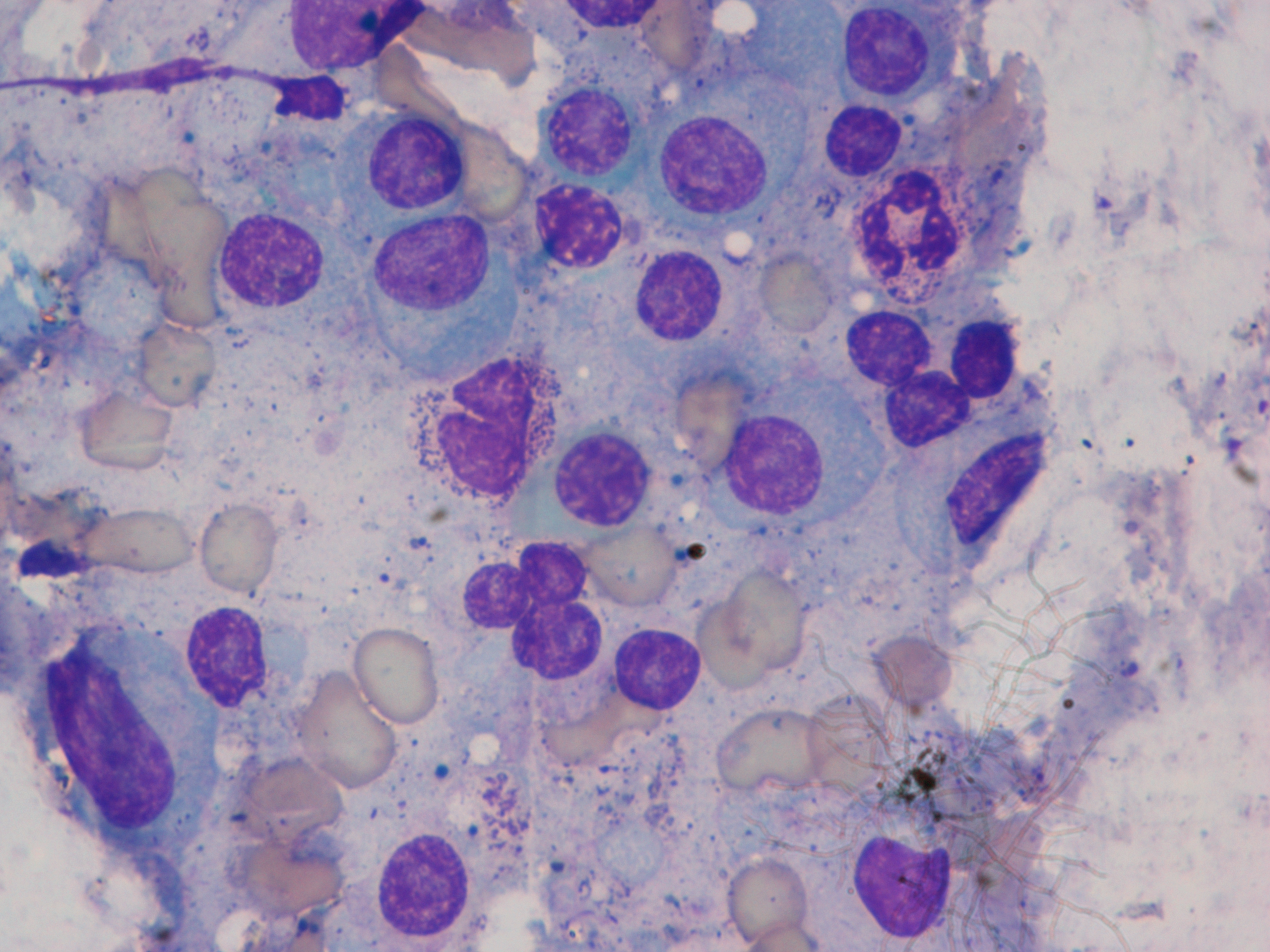
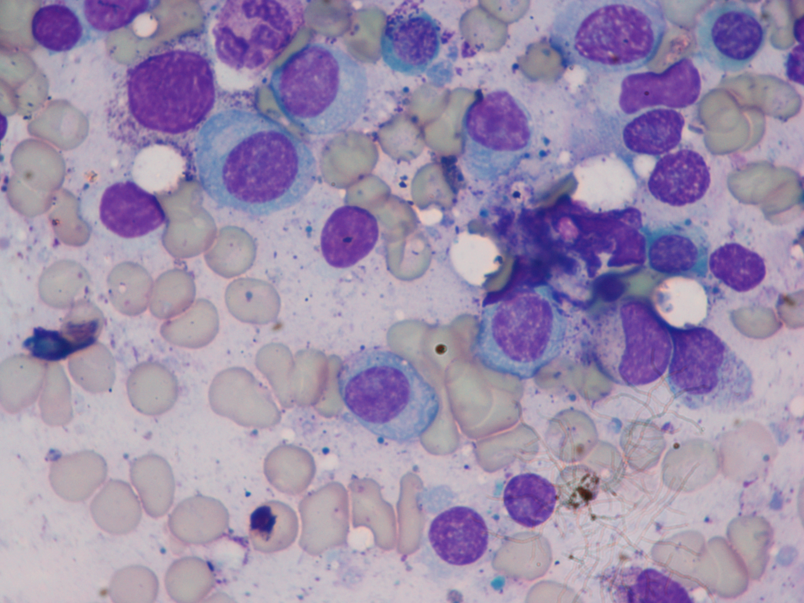
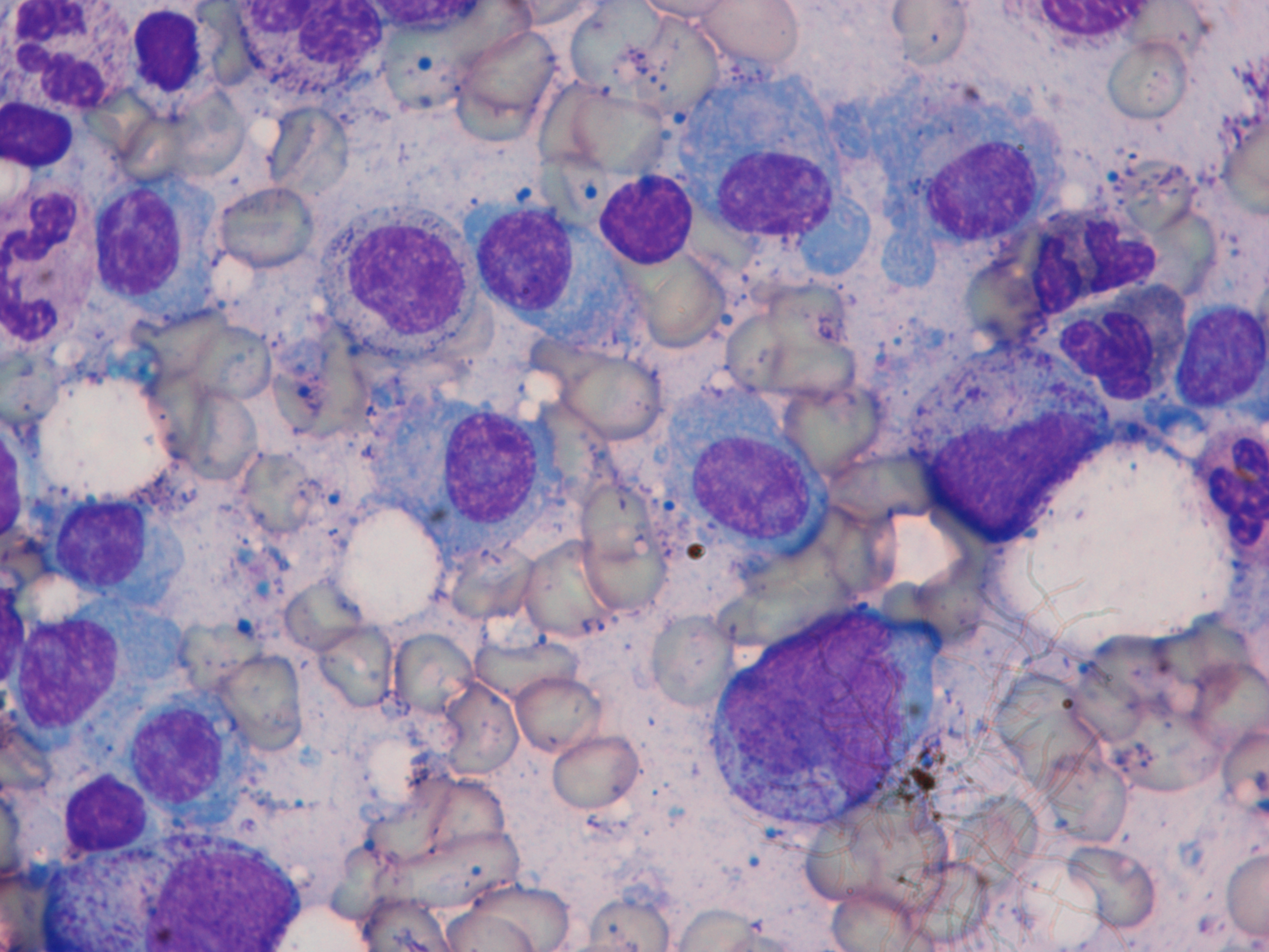
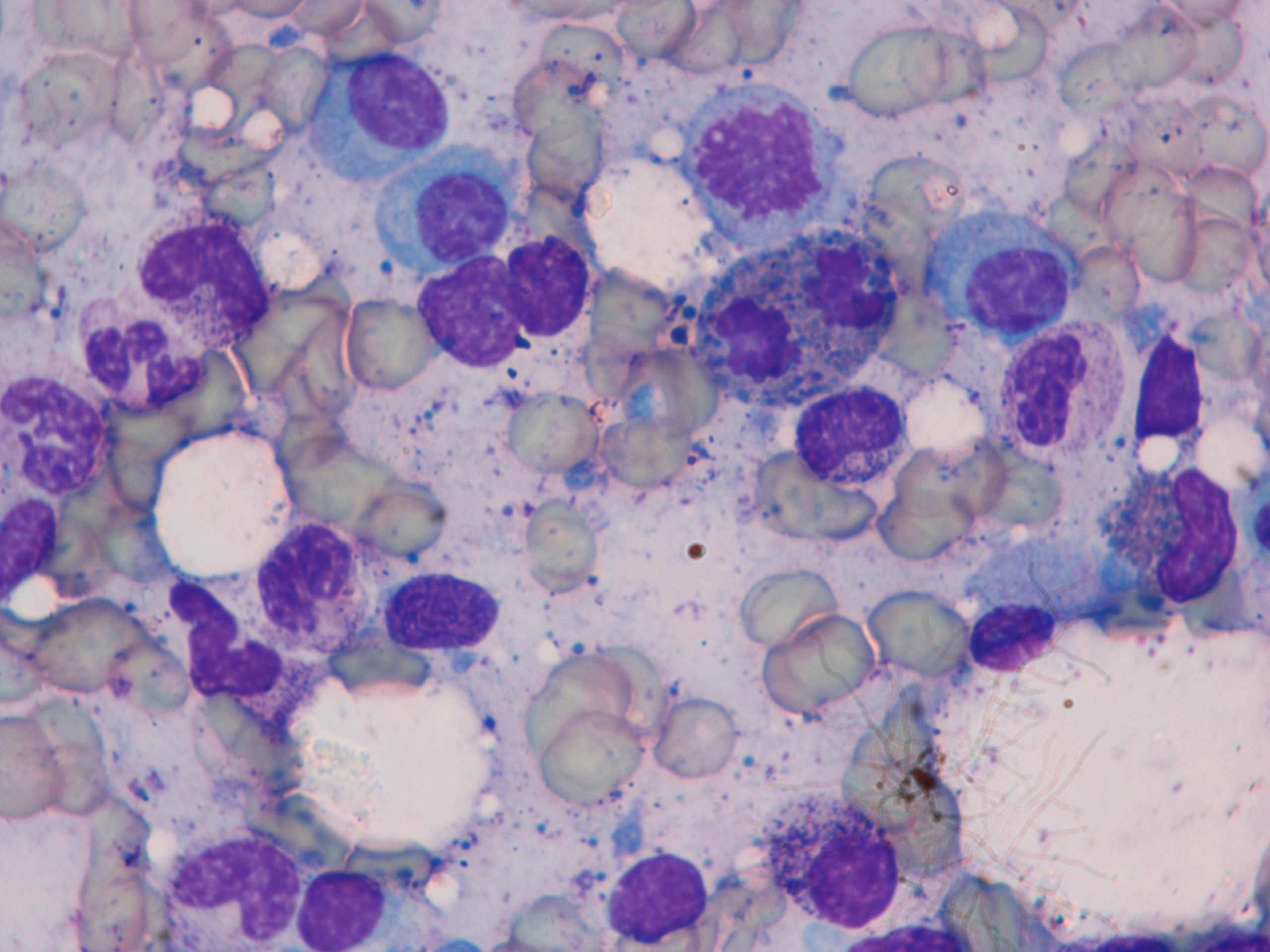
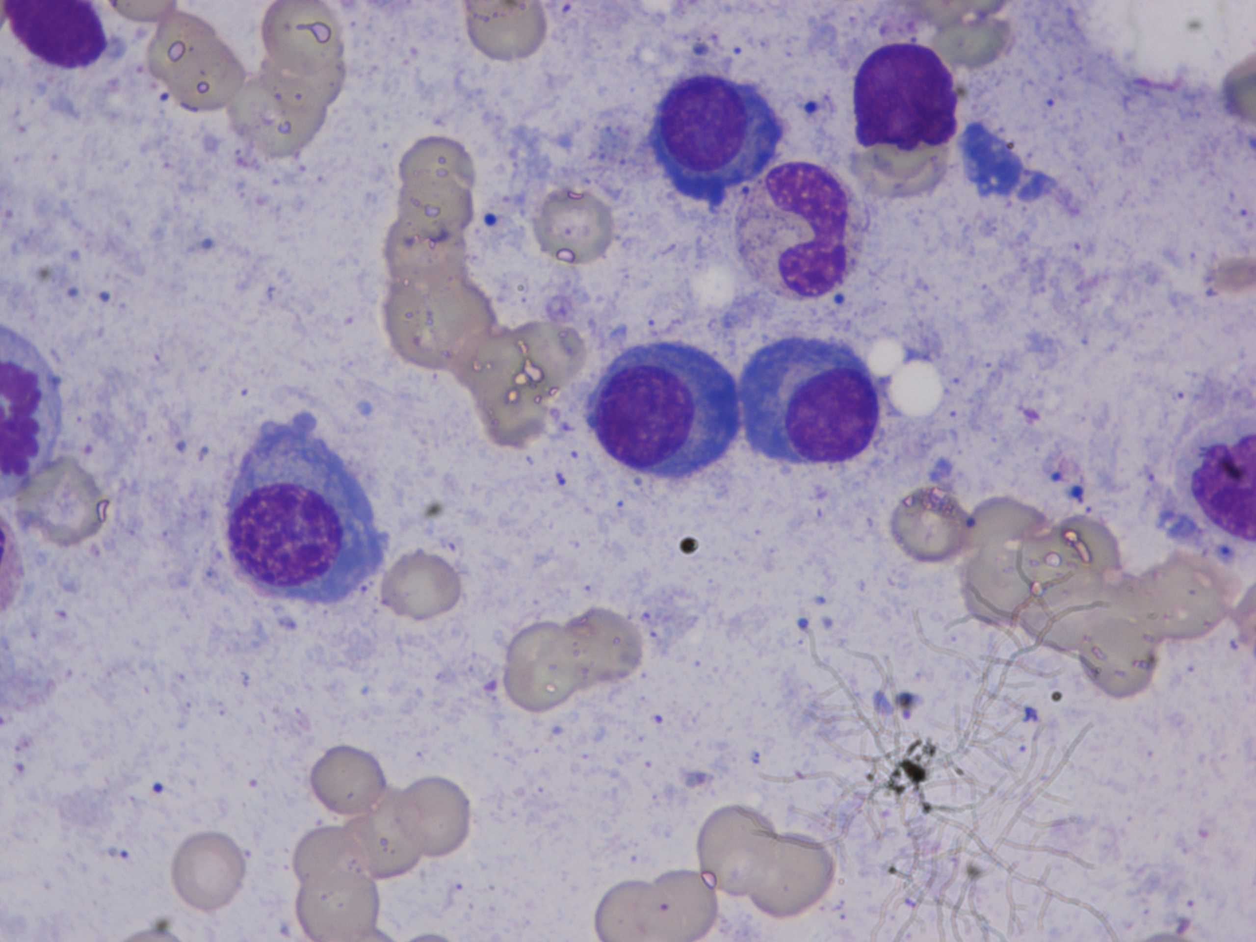
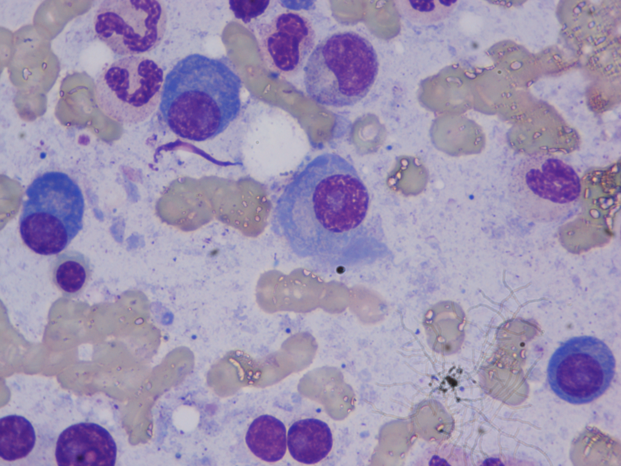
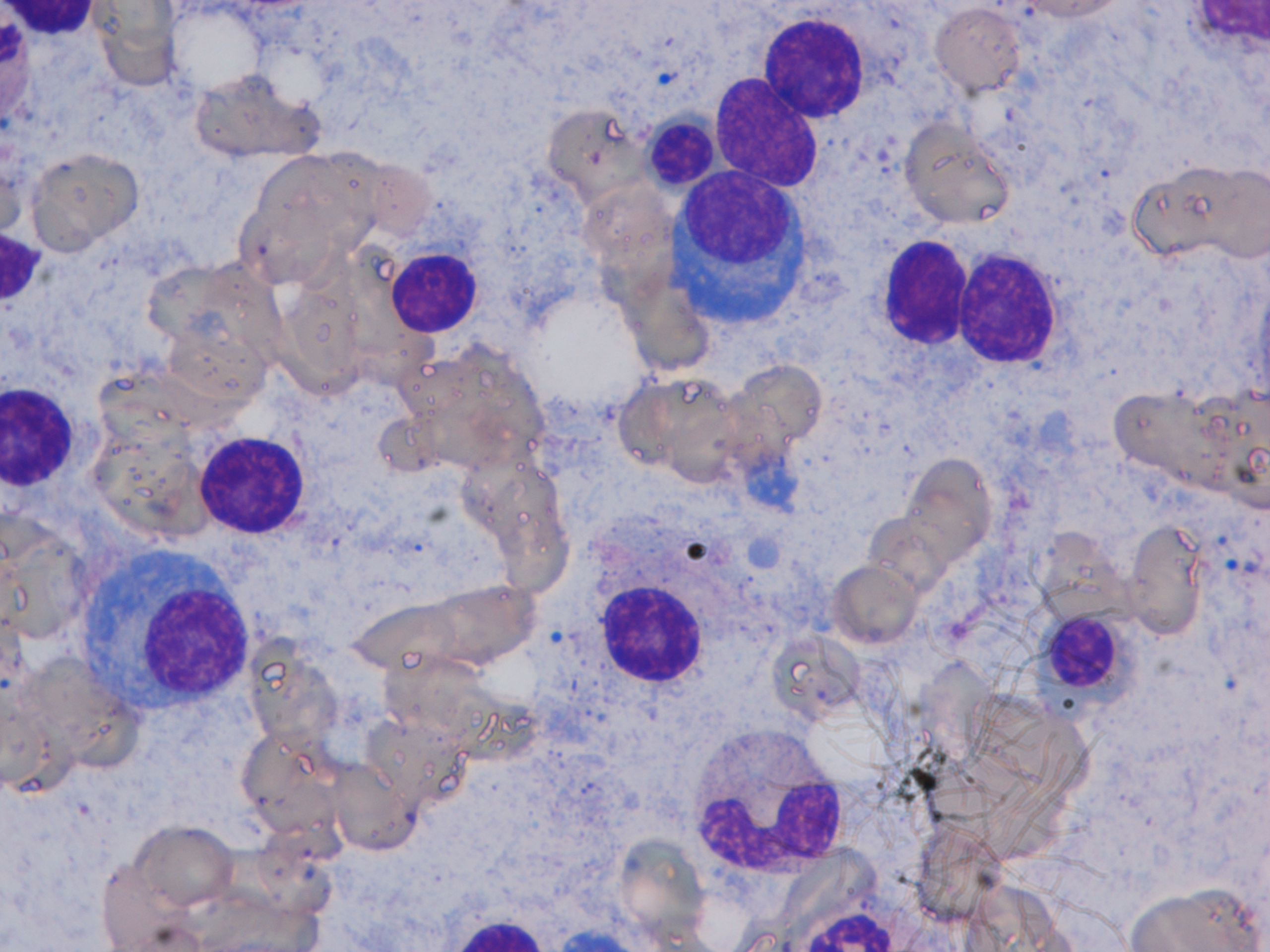
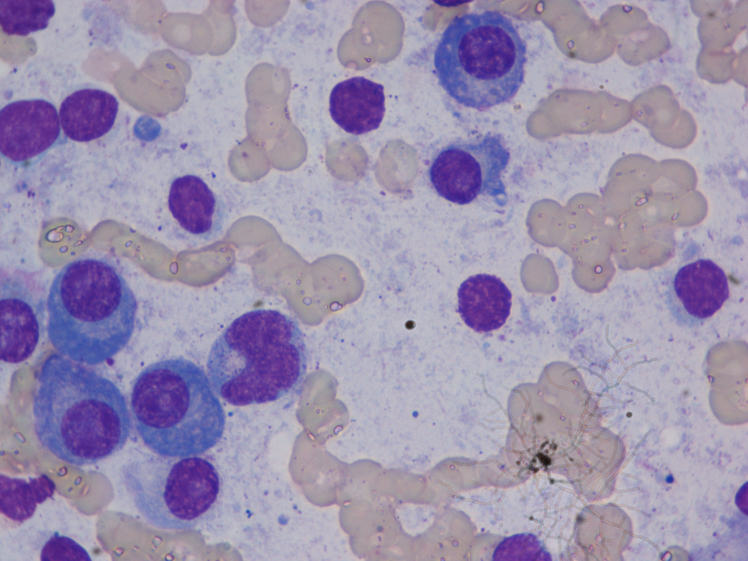
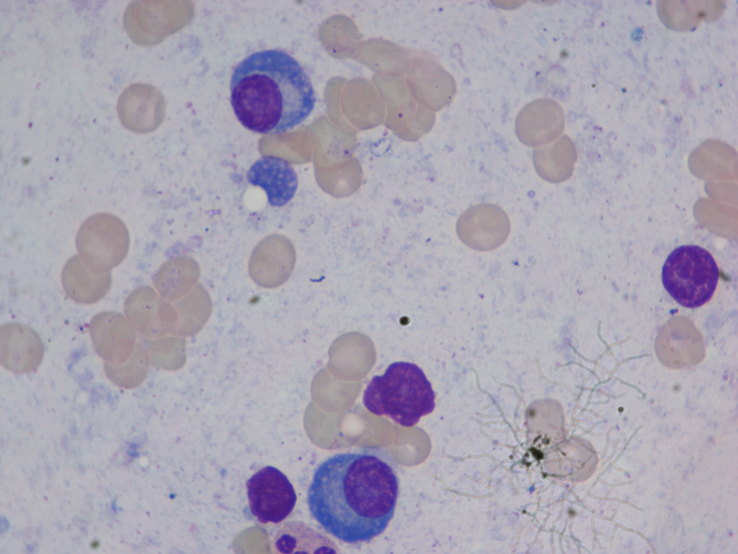
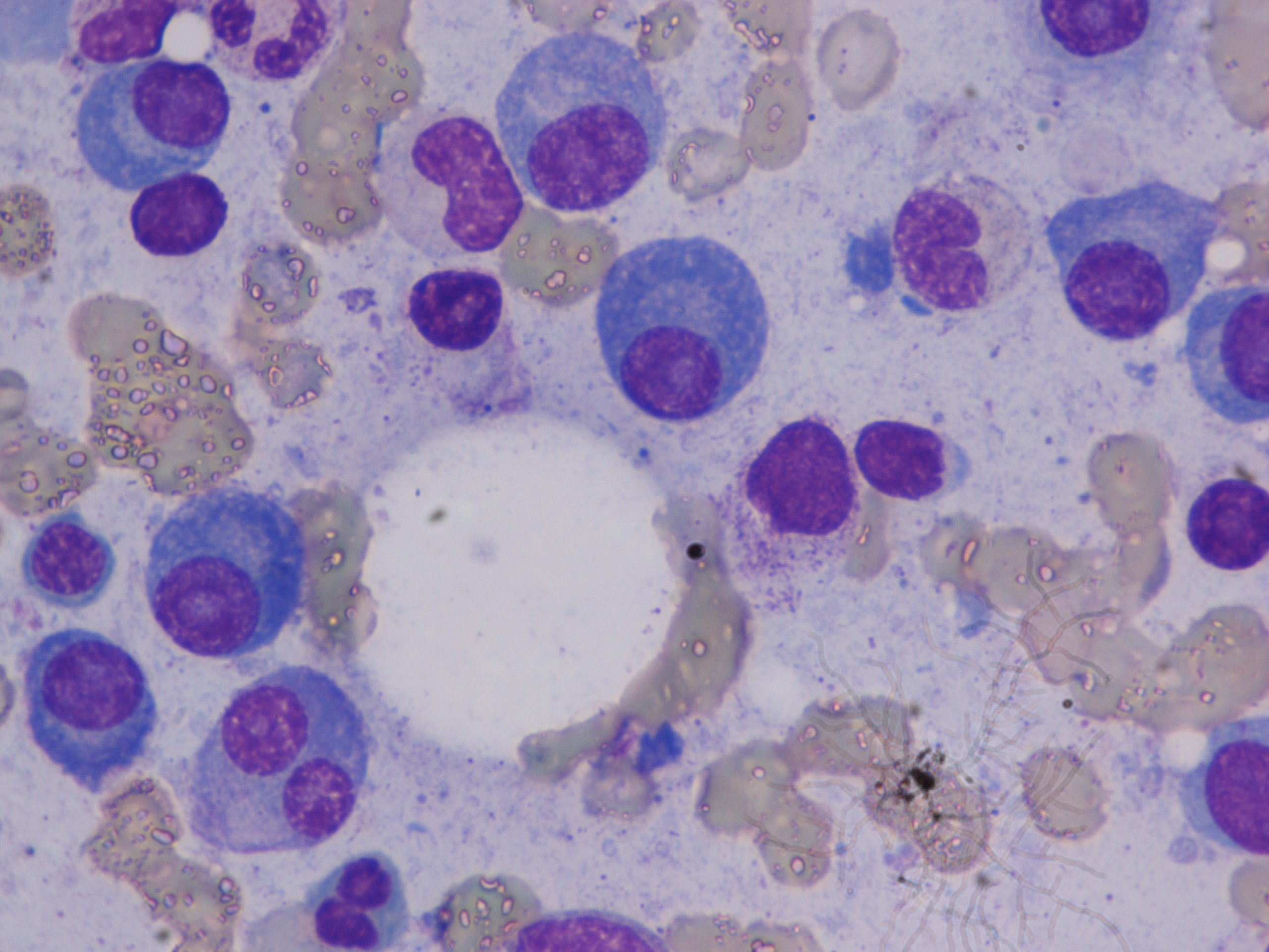
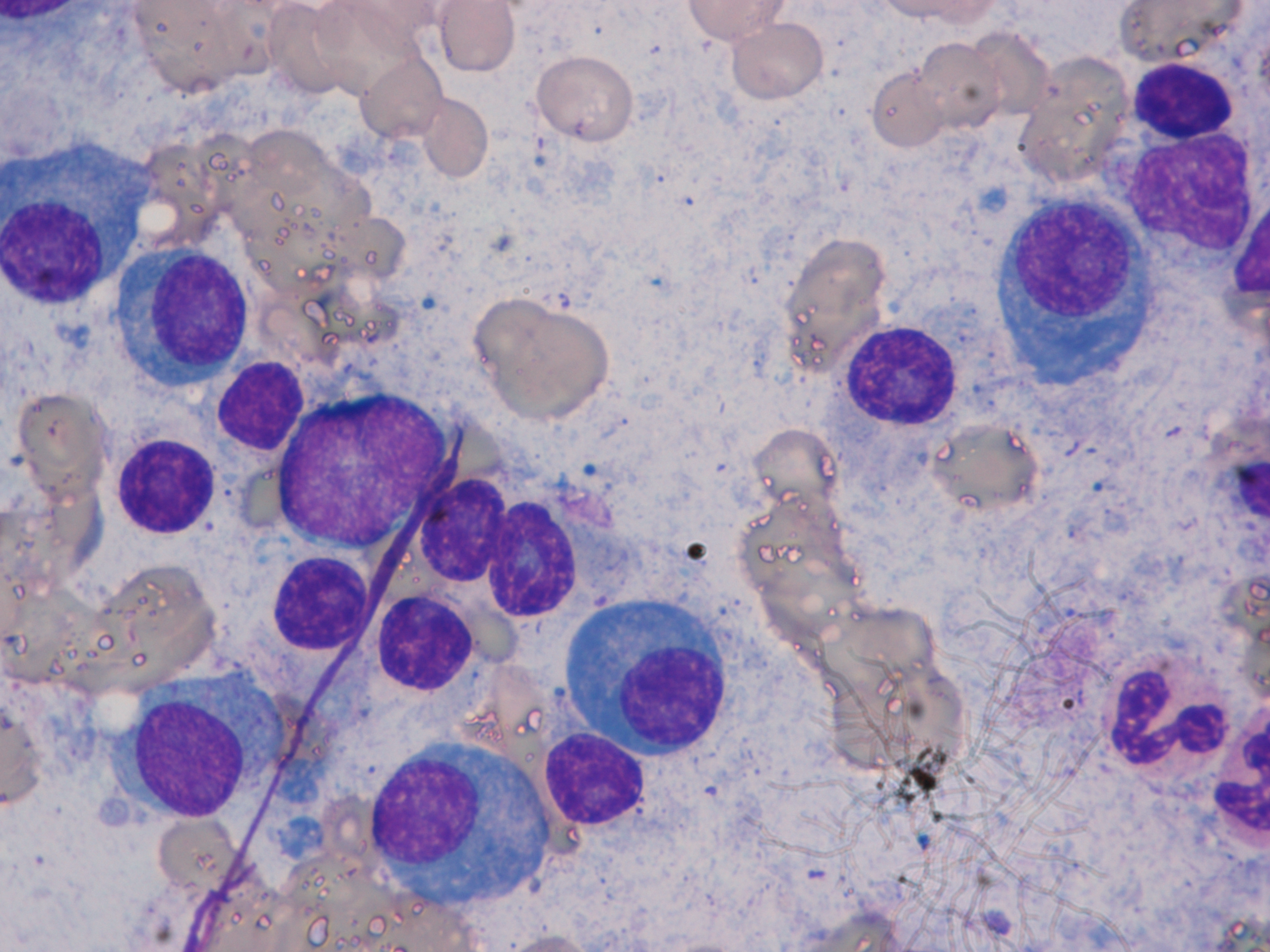
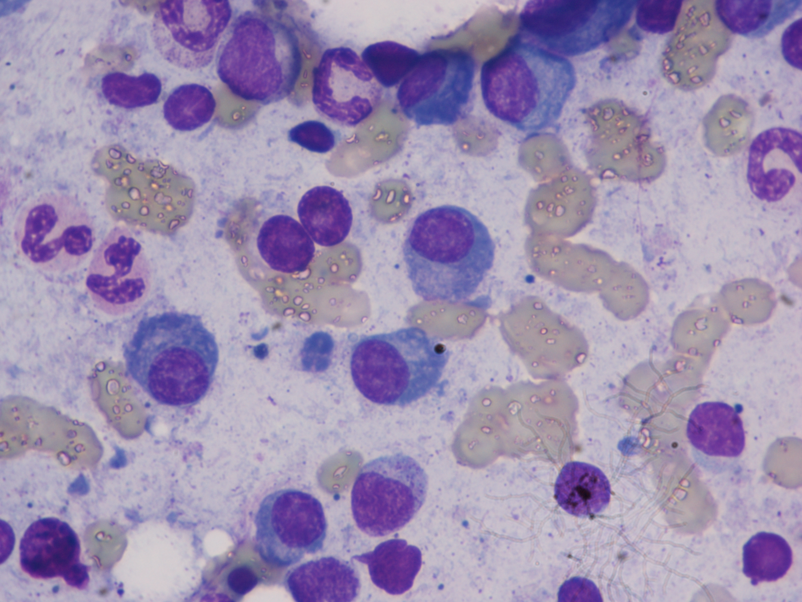
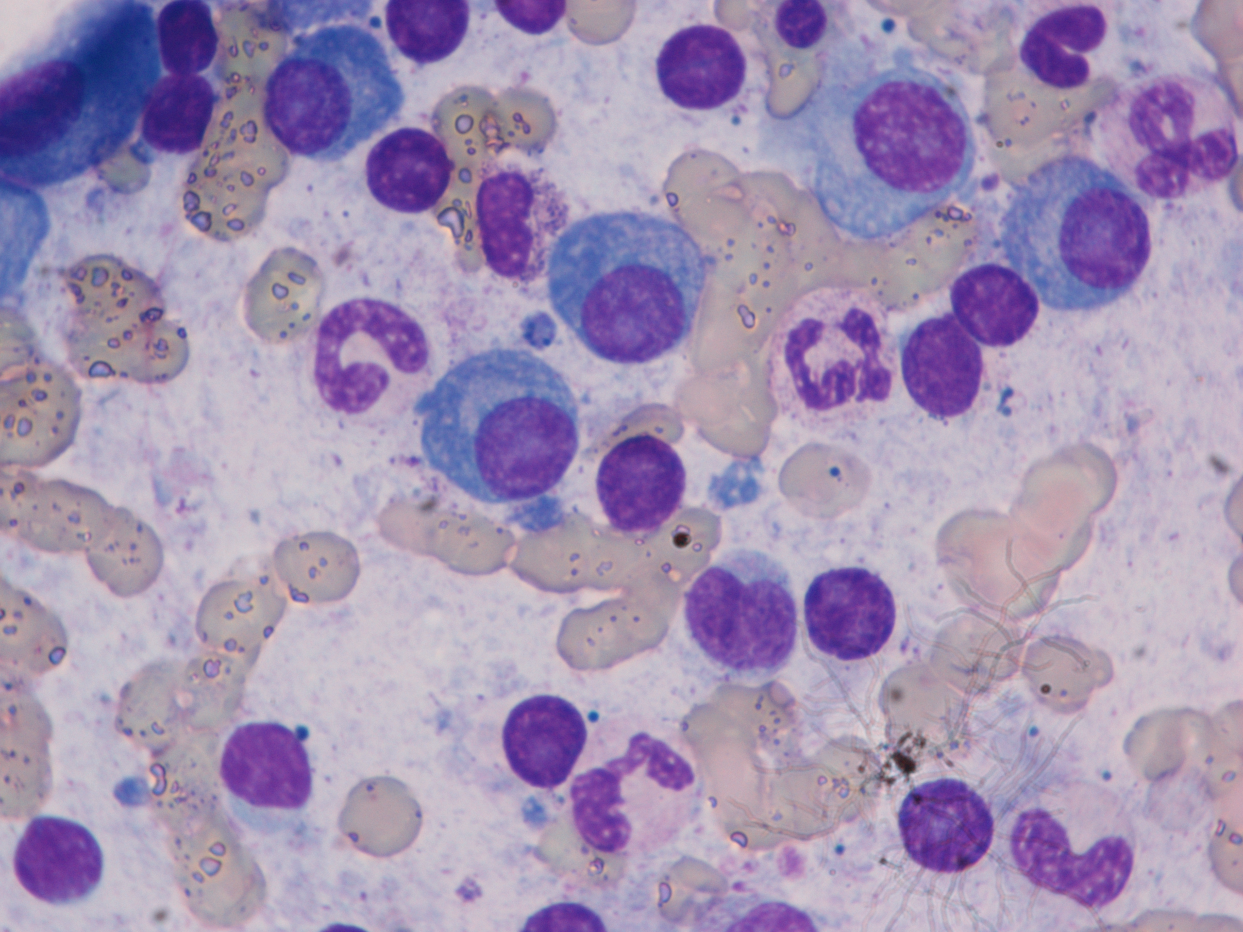
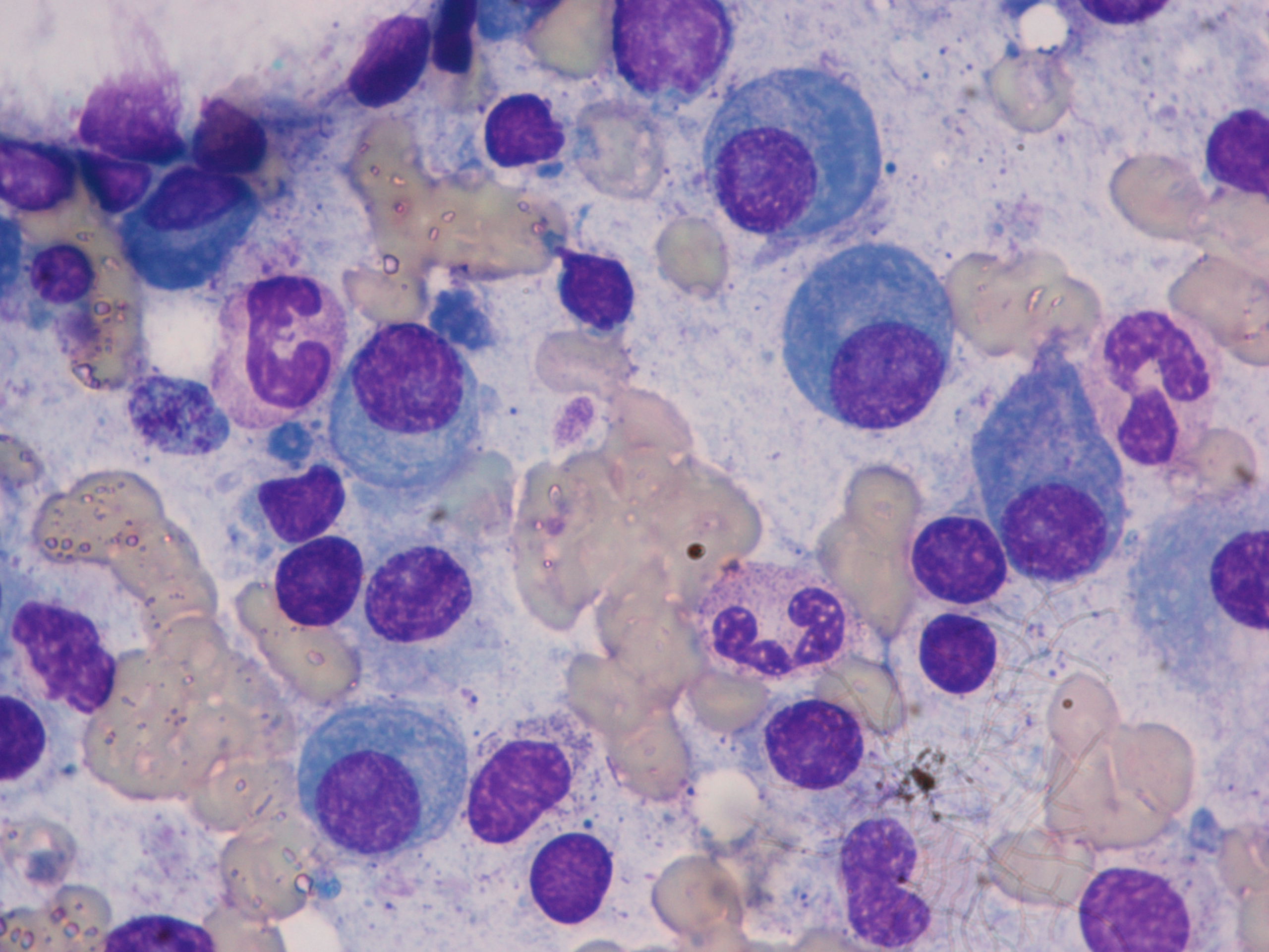
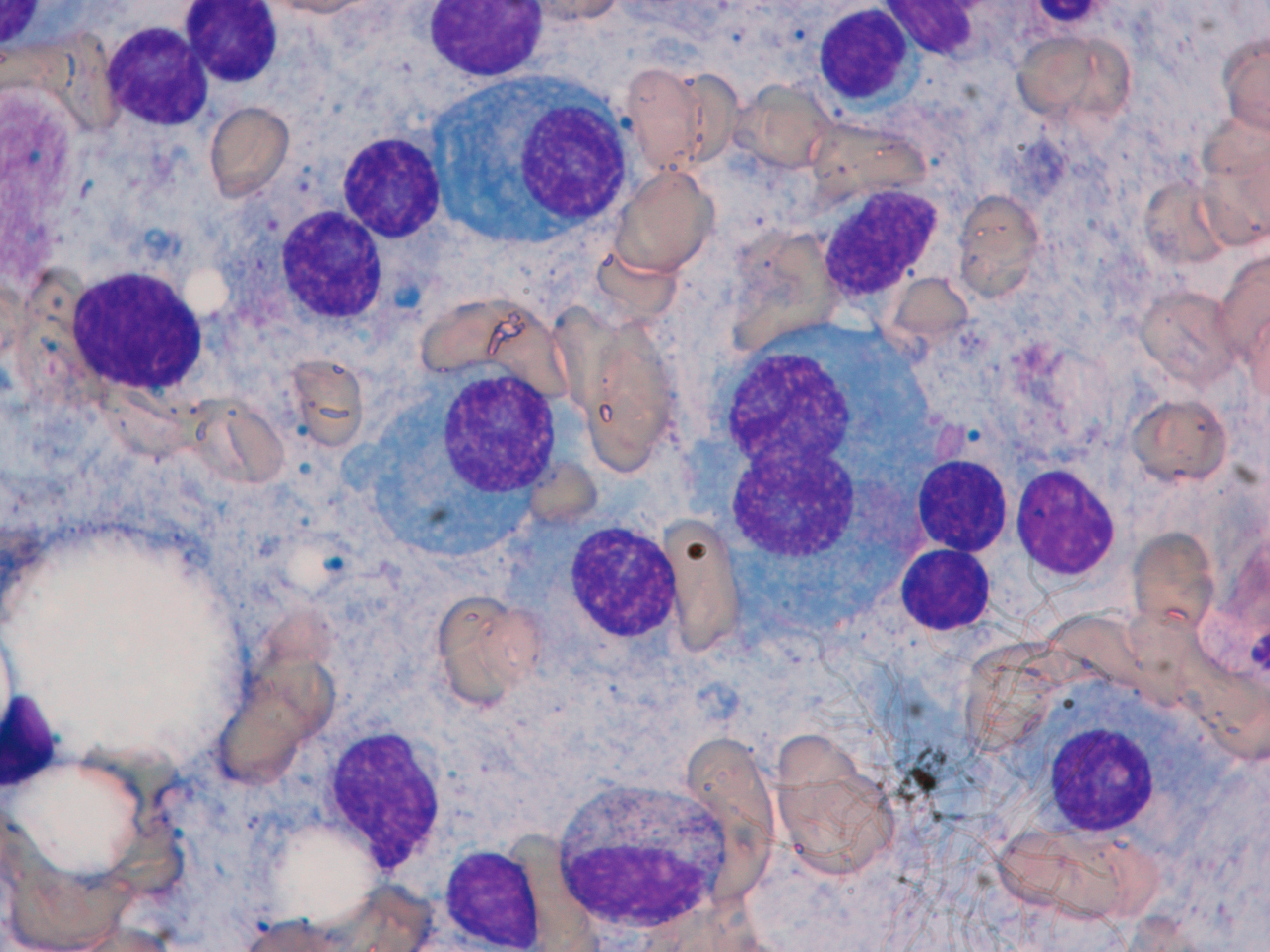
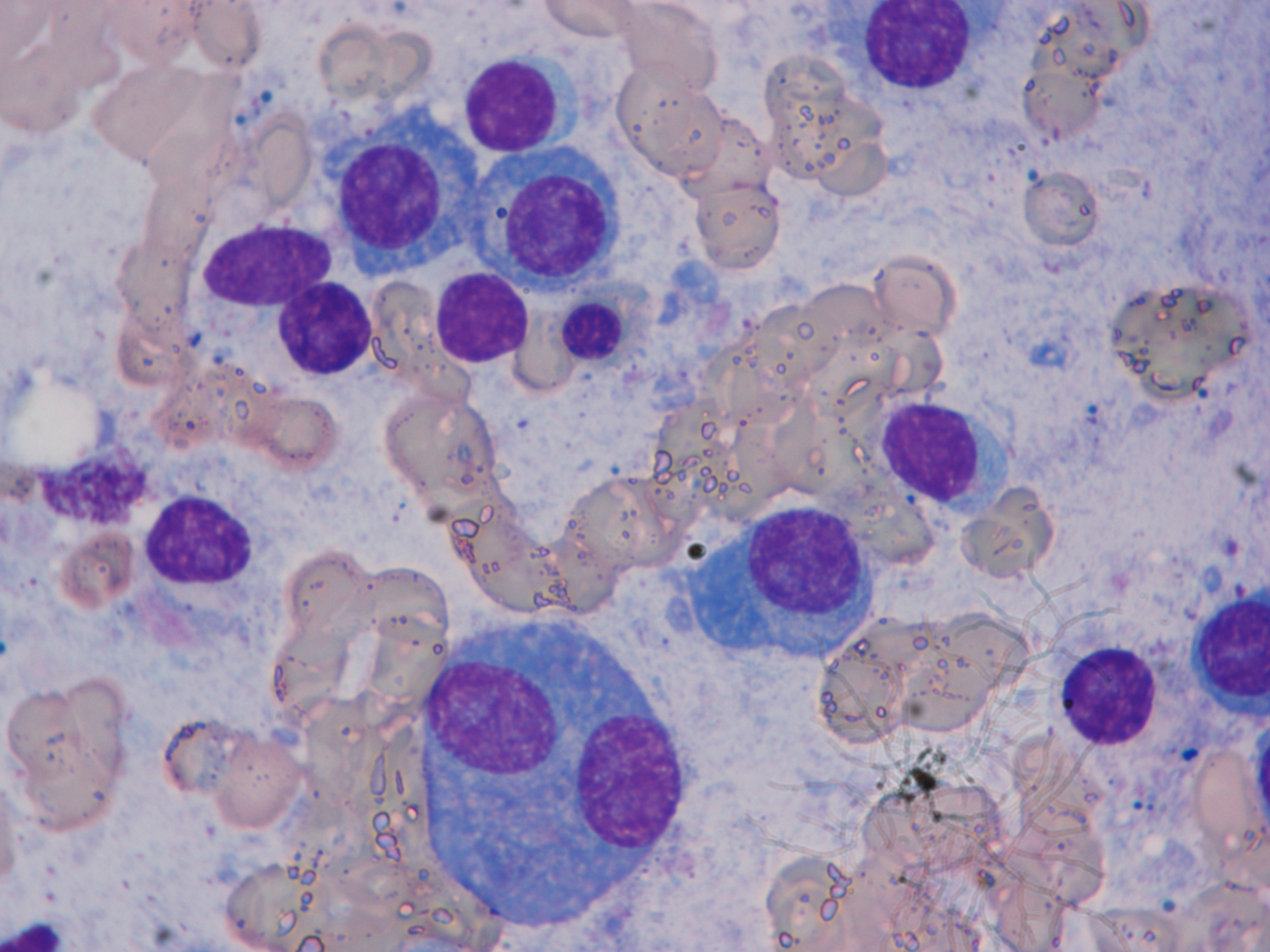
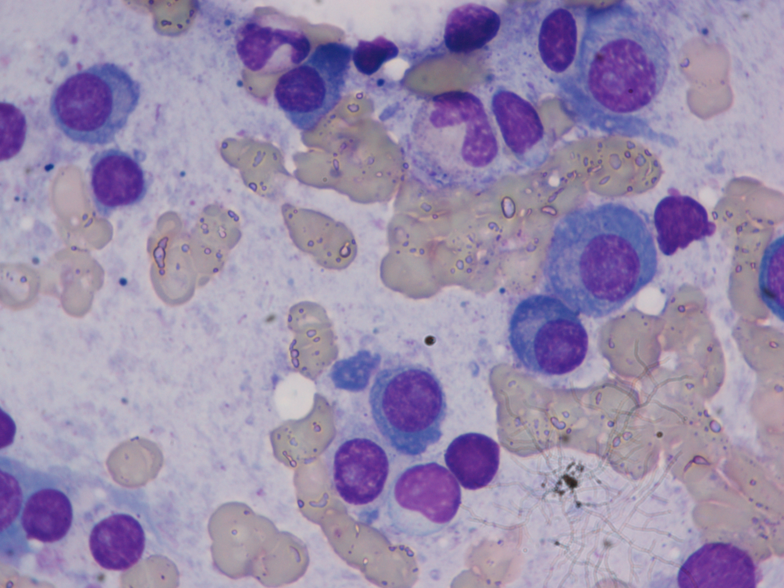
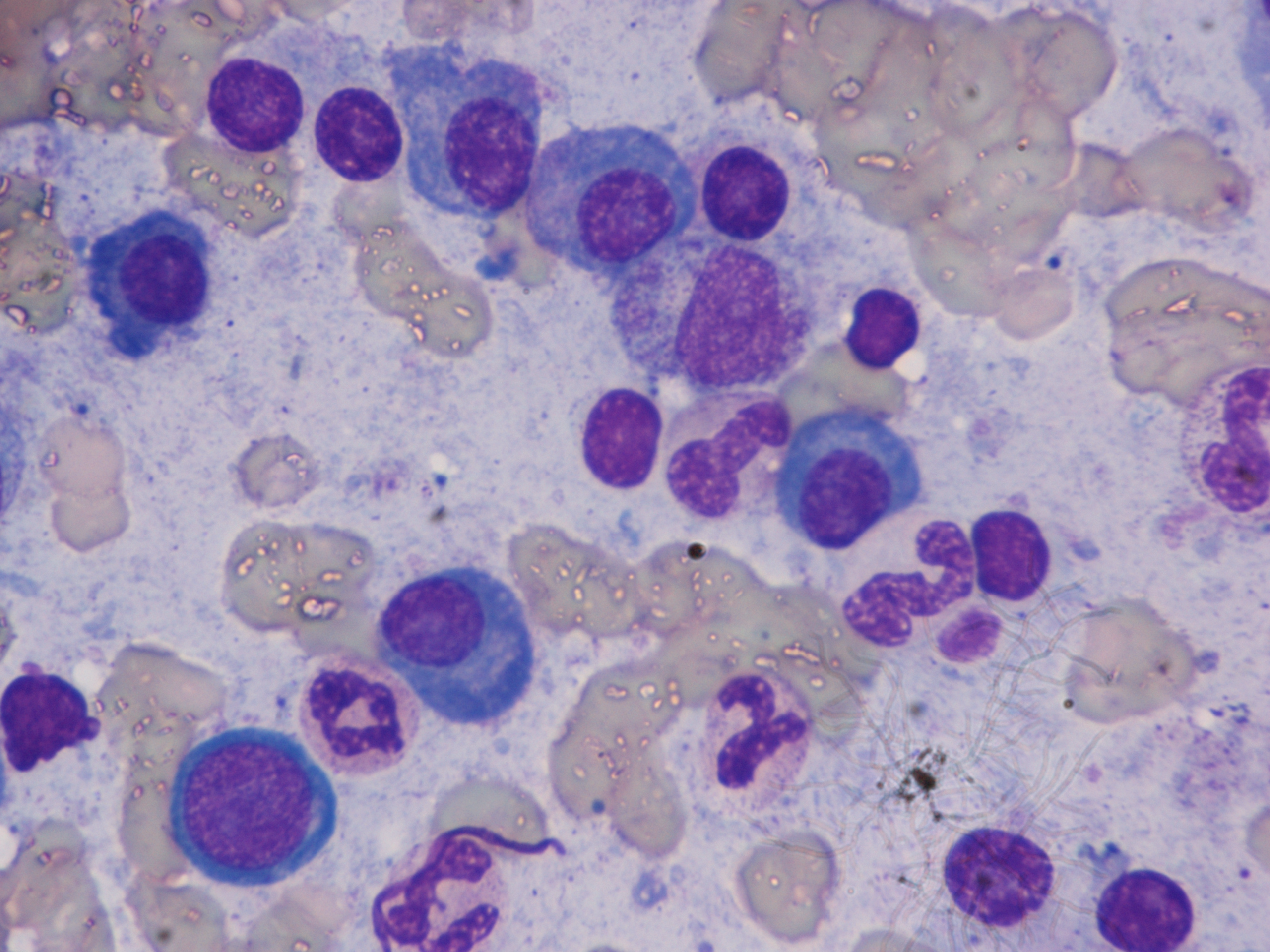
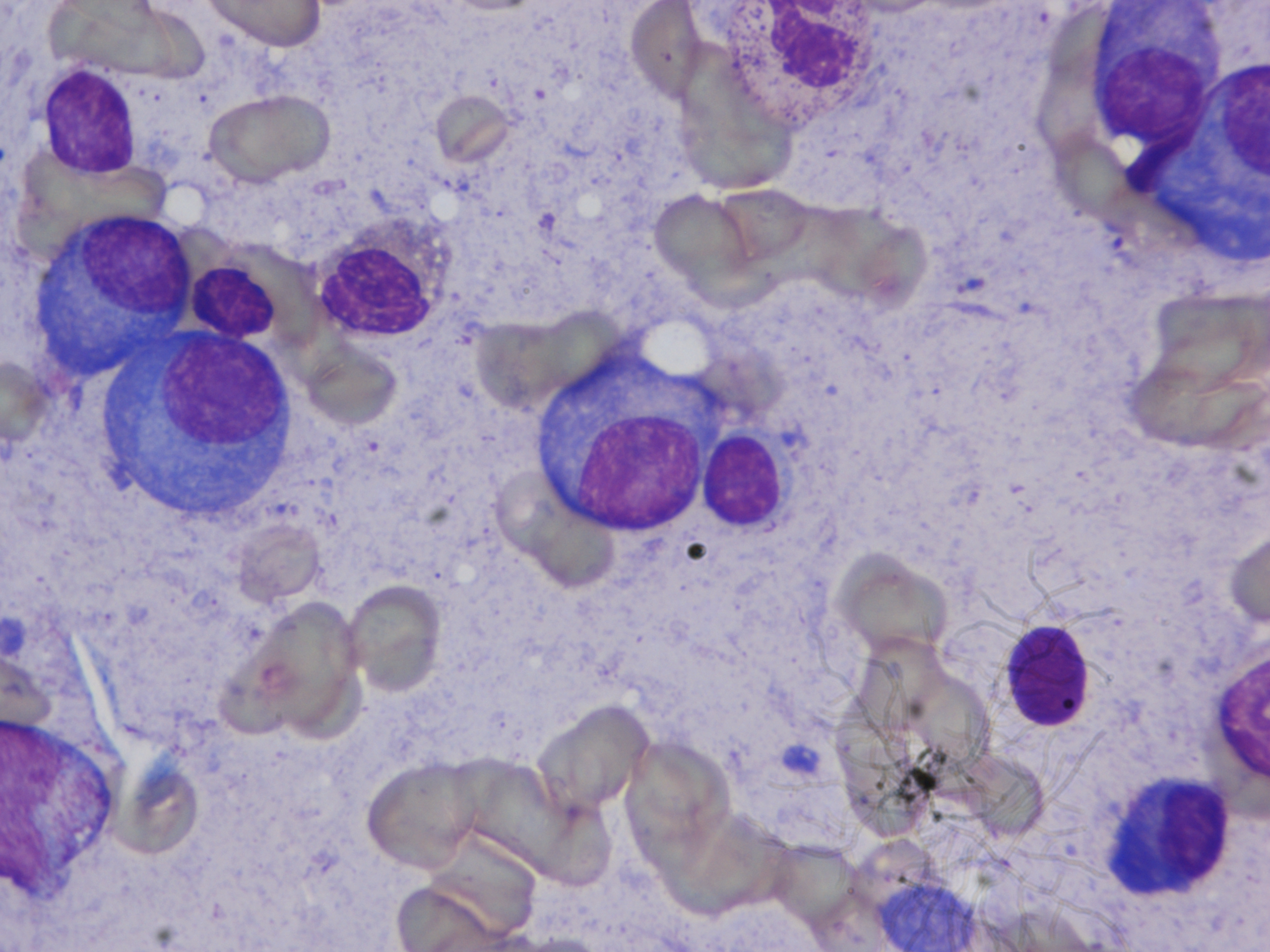
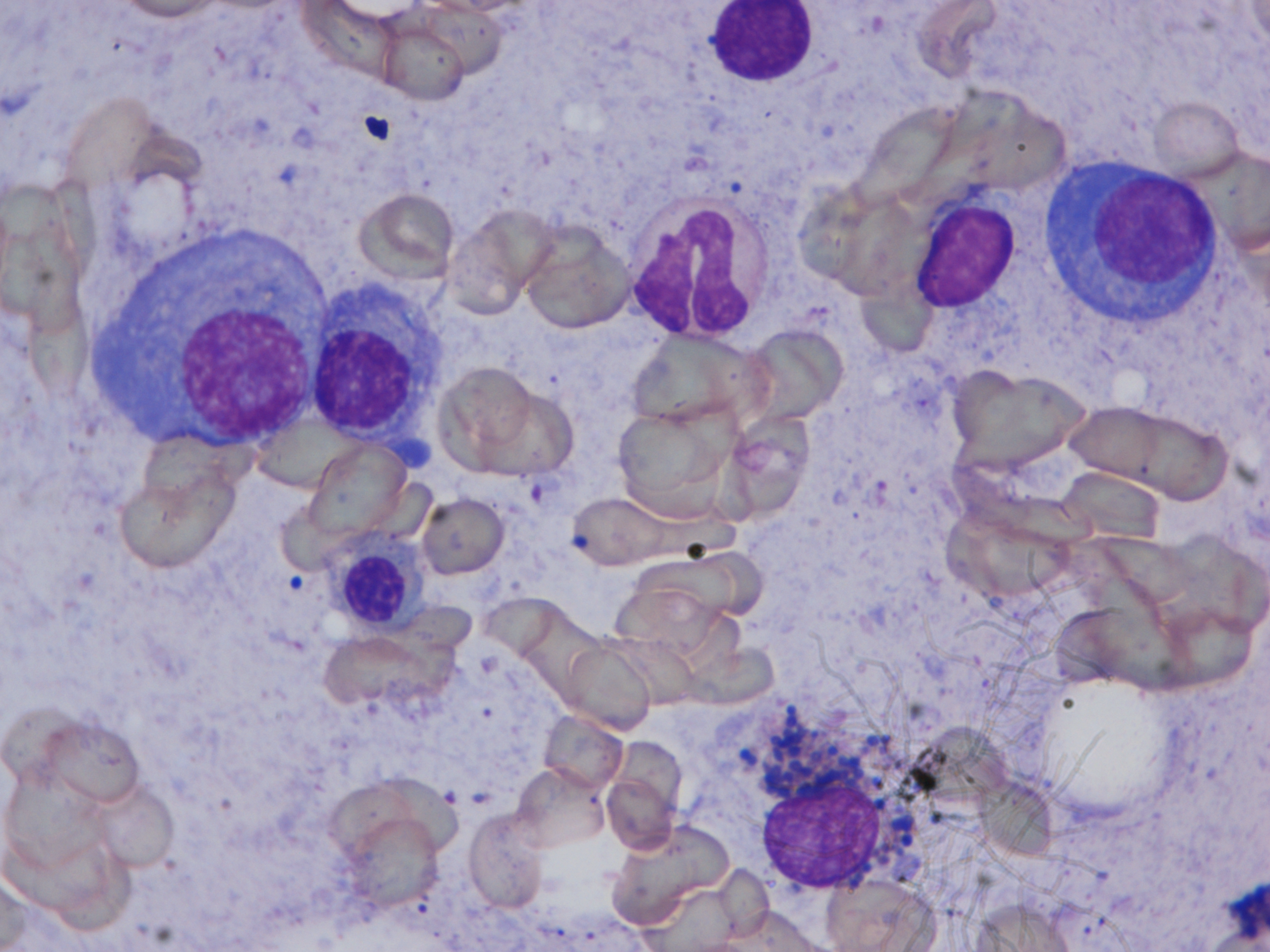
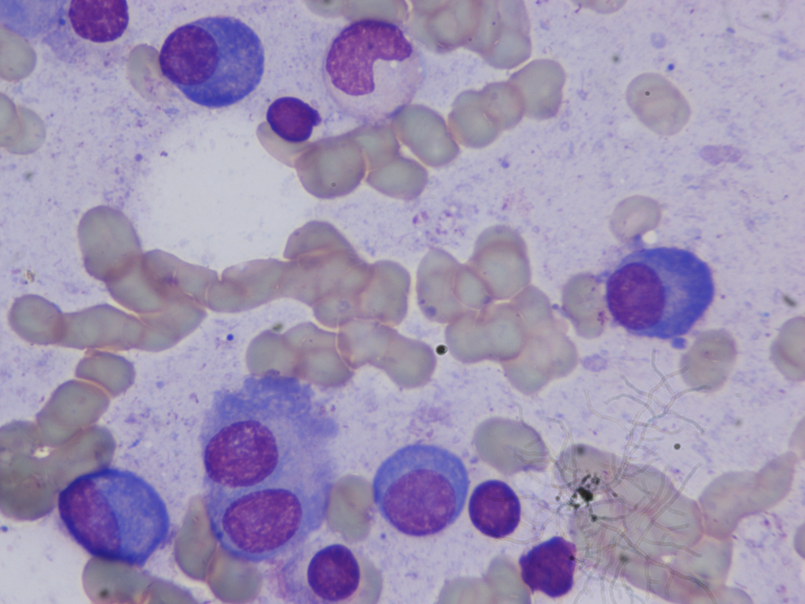
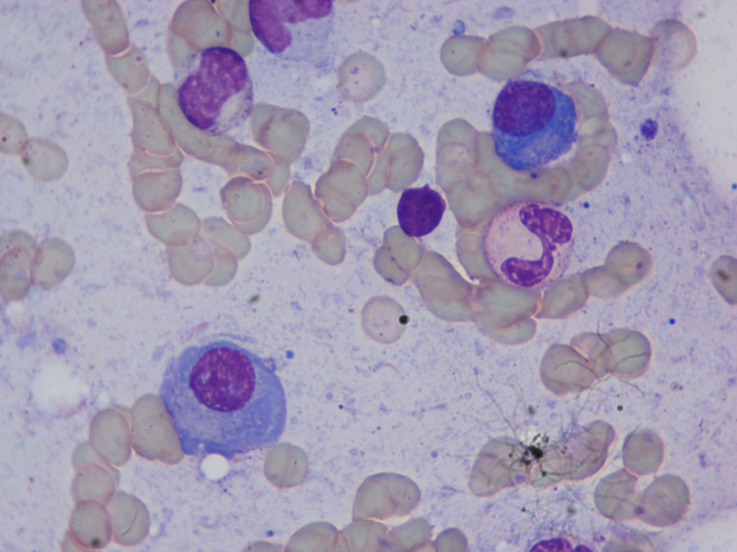
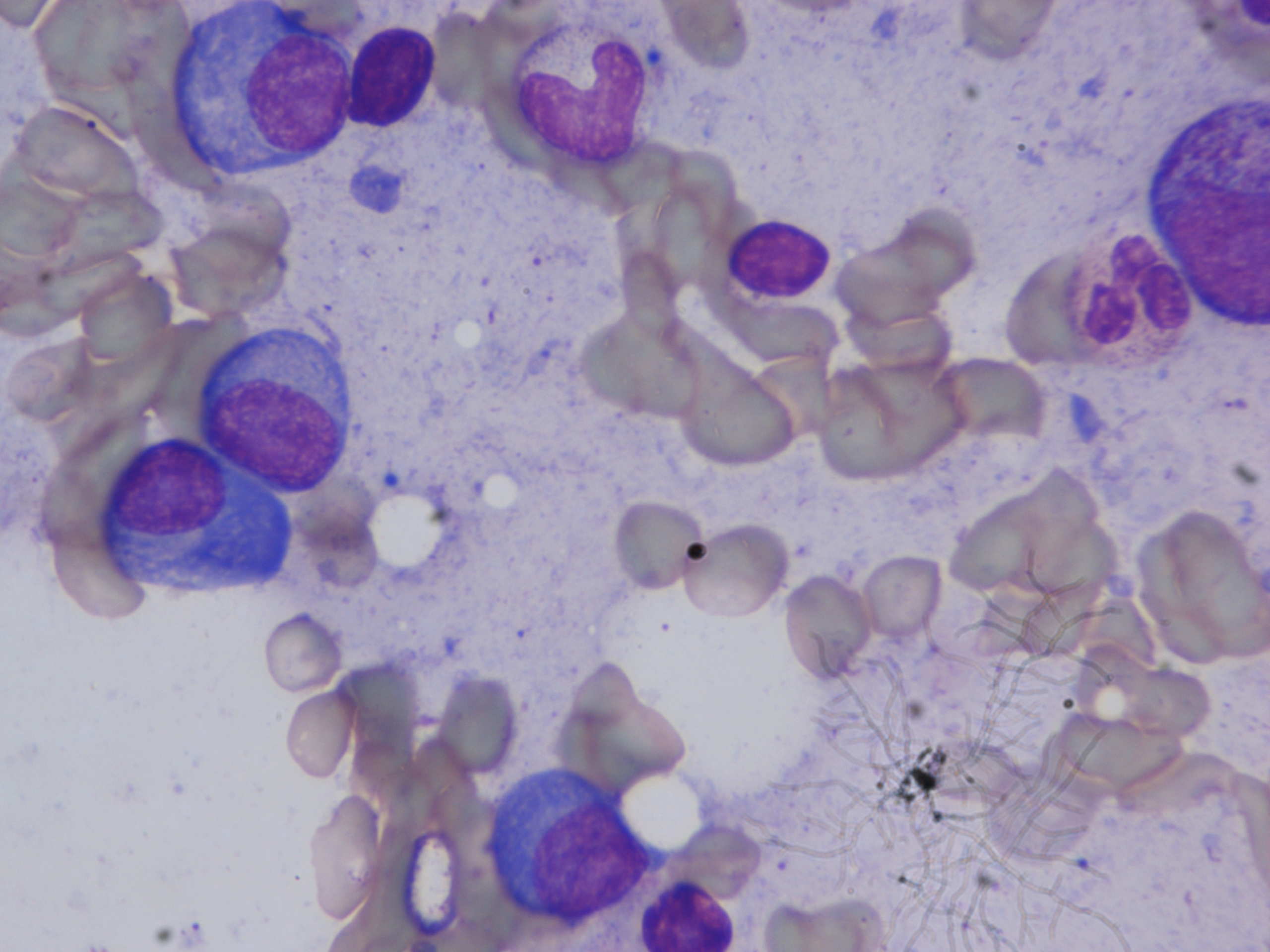
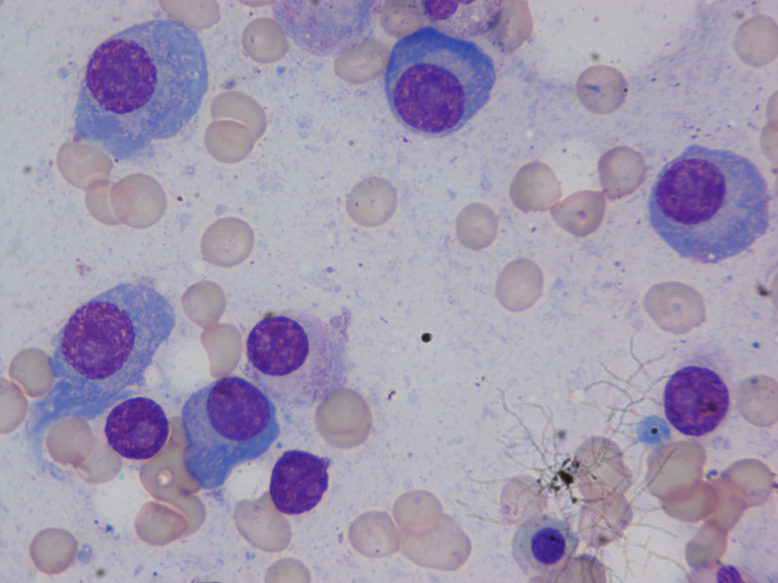
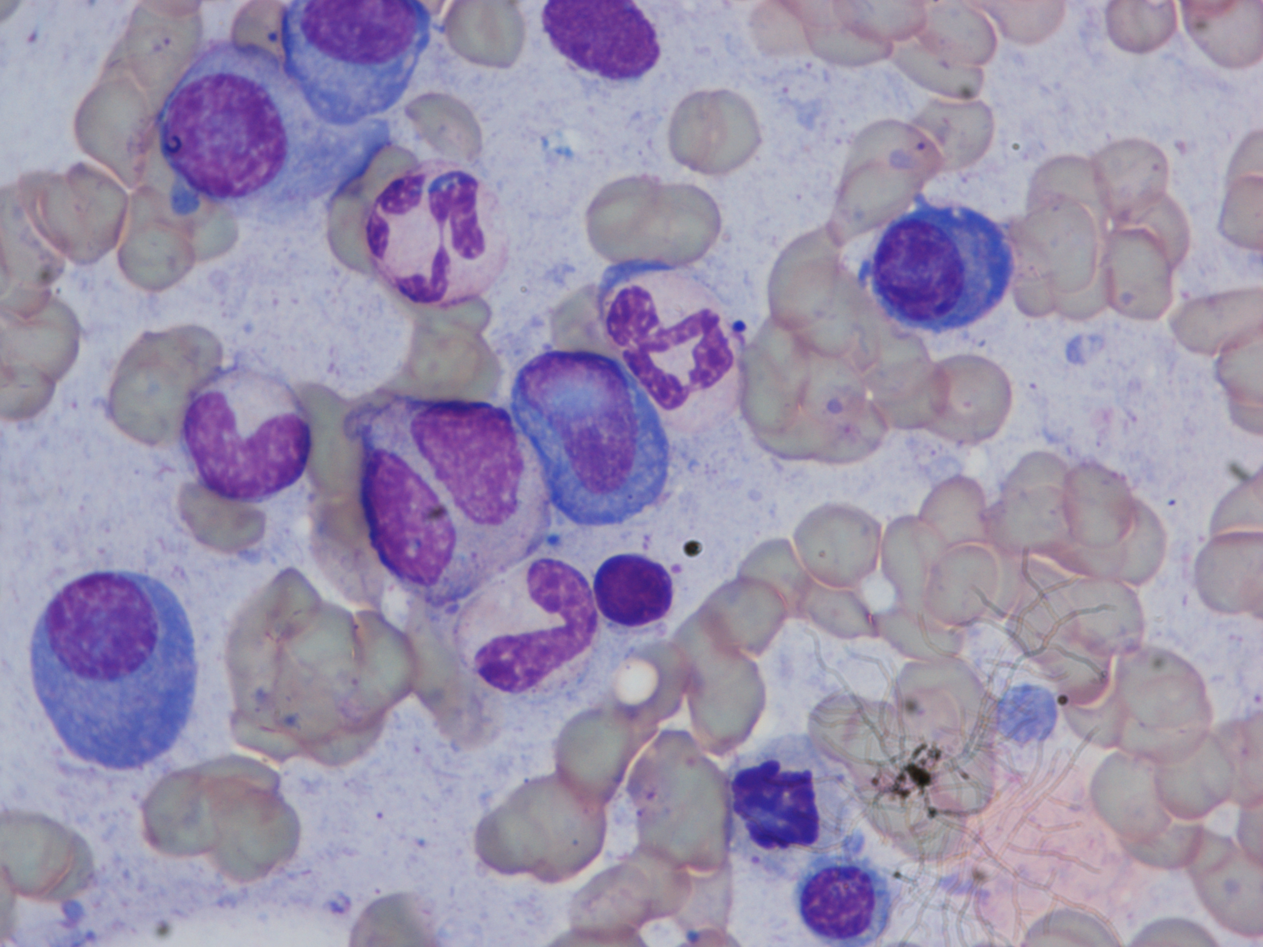
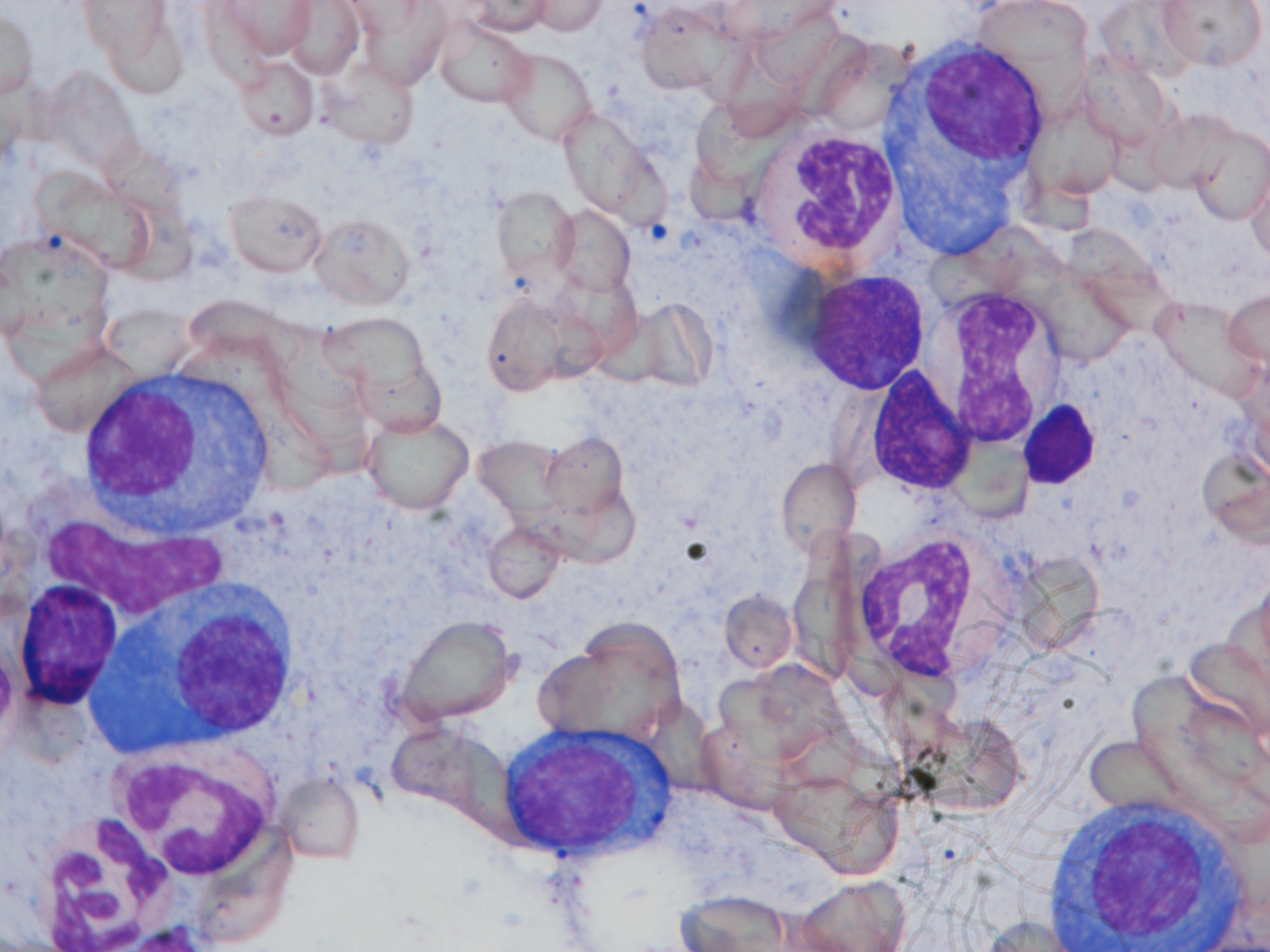
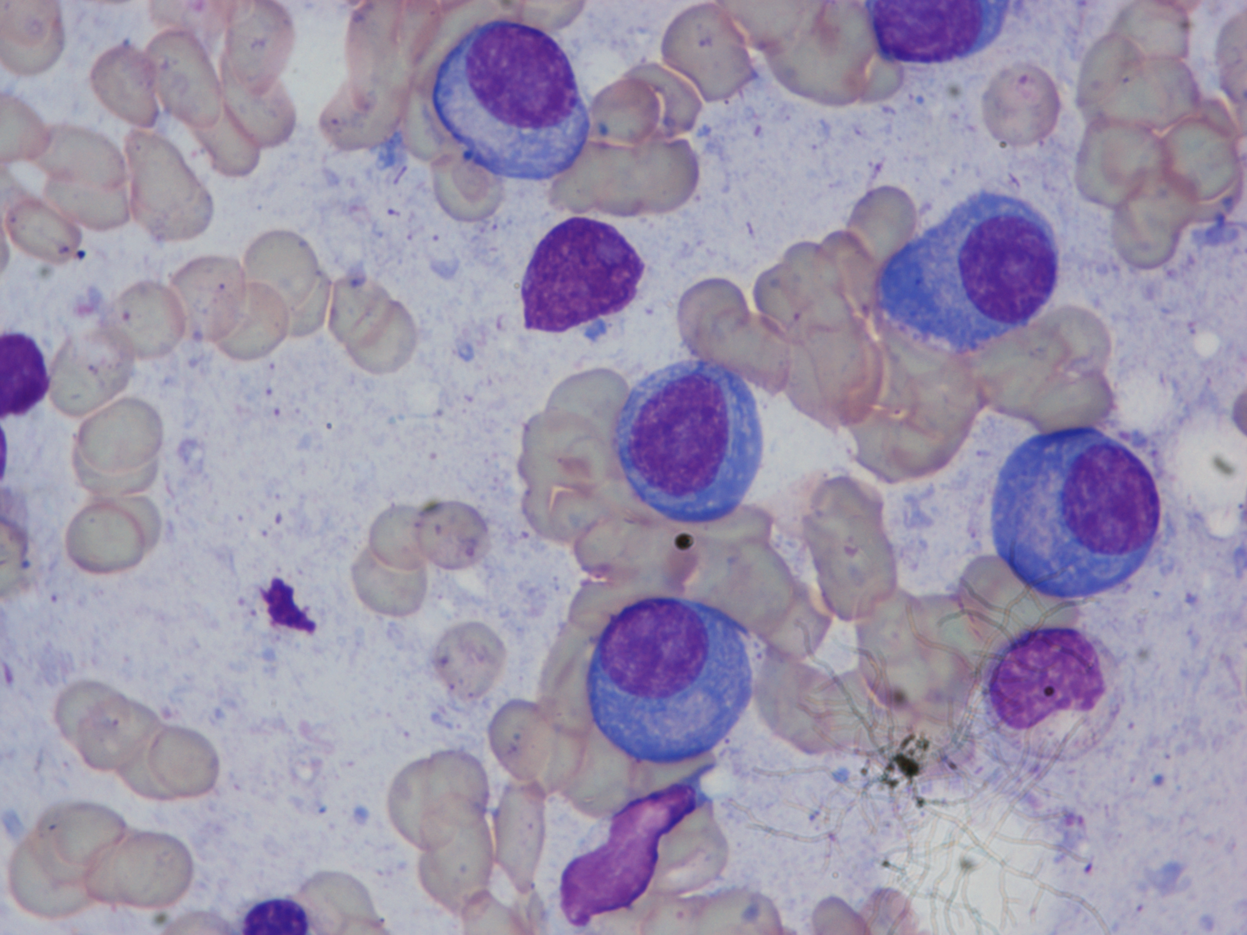
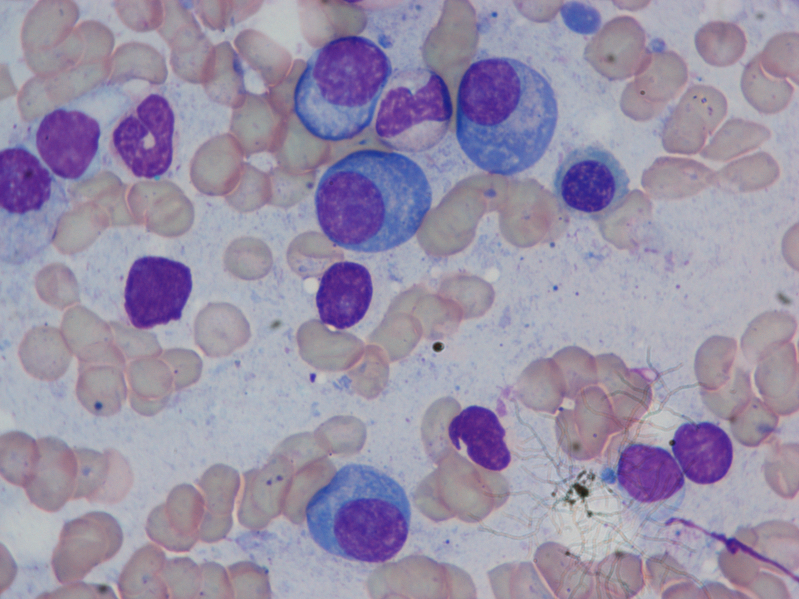
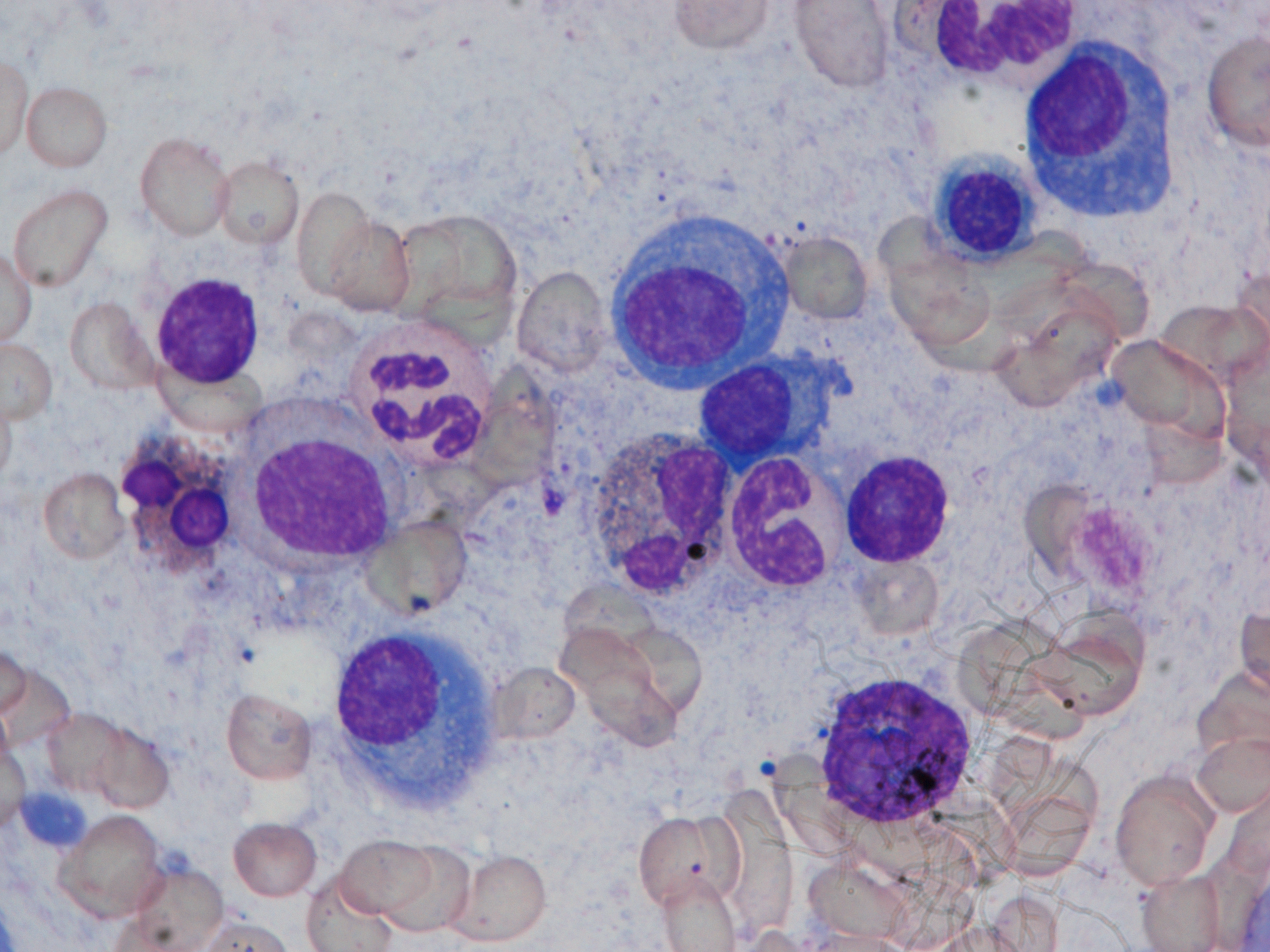
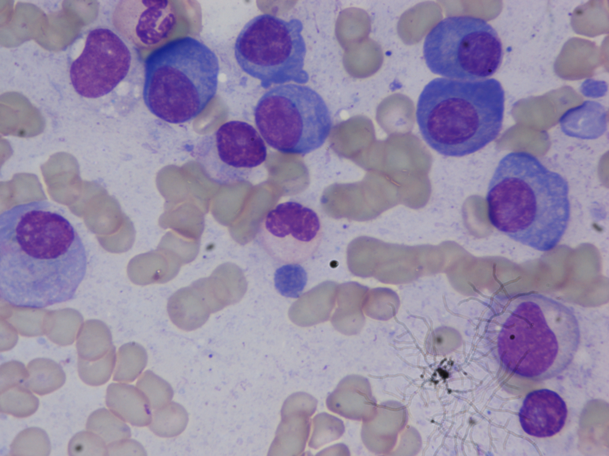
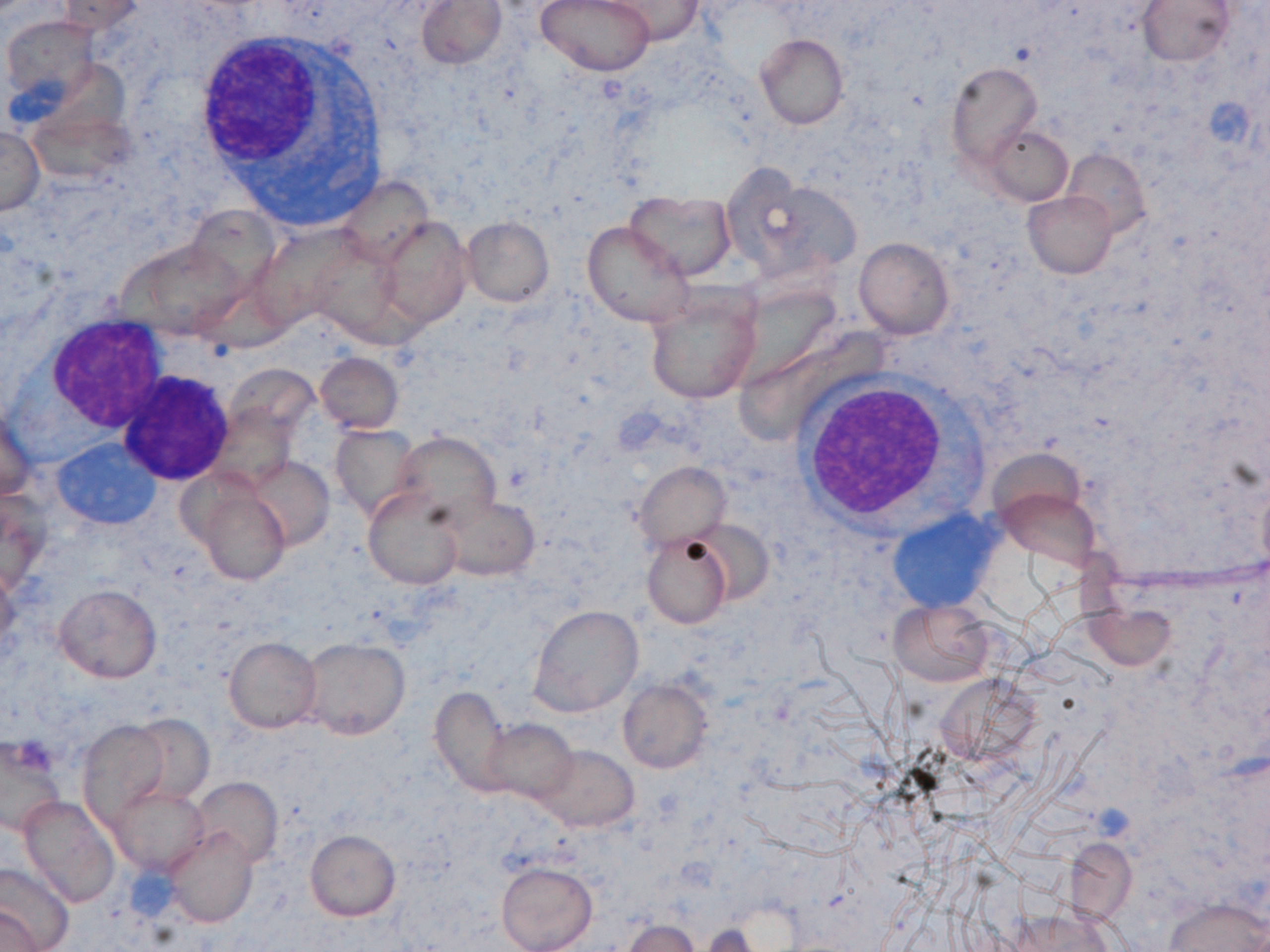
| Description: | Plasma cell segmentation is the first stage of a computer assisted automated diagnostic tool for multiple myeloma (MM). Owing to large variability in biological cell types, a method for one cell type cannot be applied directly on the other cell types. In this study, PCSeg Tool for plasma cell segmentation from microscopic medical images has presented. These images were captured from bone marrow aspirate slides of patients with multiple myeloma as per the standard guidelines. PCSeg has a robust pipeline consisting of a pre-processing step, the proposed modified multiphase level set method followed by post-processing steps including the watershed and circular Hough transform to segment clusters of cells of interest and to remove unwanted cells. Our modified level set method utilizes prior information about the probability densities of regions of interest (ROIs) in the color spaces and provides a solution to the minimal-partition problem to segment ROIs in one of the level sets of a two-phase level set formulation. PCSeg tool is tested on a number of microscopic images and provides good segmentation results on single cells as well as efficient segmentation of plasma cell clusters. |
| Publications: | https://doi.org/10.1371/journal.pone.0207908 |
| Funding agency: | Department of Science and Technology, Govt. of India and Ministry of Communication and IT, Govt. of India. |
| Grant Number: | EMR/2016/006183 and 1(7)2014-ME&HI |
| Ethics Statement: | Download |
| Any Other Information : | The original version of this dataset is available at The Cancer Imaging Archive (TCIA; https://www.cancerimagingarchive.net/collection/mimm_sbilab/). The TCIA citation is: Gupta, R., & Gupta, A. (2019). MiMM_SBILab Dataset: Microscopic Images of Multiple Myeloma [Data set]. The Cancer Imaging Archive. https://doi.org/10.7937/tcia.2019.pnn6aypl. This collection has also been uploaded to the Harvard Blood Cancer Dataverse website. Please refer to DOI: https://doi.org/10.7910/DVN/XCX7ST for more information. |
| Additional File: | Download |
| Acknowledgments: | Authors gratefully acknowledge the research funding support (Grant Number: 1(7)/2014-ME&HI) from the Ministry of Communication and IT, Govt. of India and funding support (Grant: EMR/2016/006183) from the Department of Science and Technology, Govt. of India for this research work. The funders had no role in study design, data collection and analysis, decision to publish, or preparation of the manuscript. We would also like to thank the support of Dr. Hossein and Mrs. Saeedizadeh for providing their code for the experiments. |
| Sr.No | First name | Last name | Organization | Designation | |
|---|---|---|---|---|---|
| 1 | Anubha | Gupta | anubha@iiitd.ac.in | SBILab, Department of ECE, Indraprastha Institute of Information Technology-Delhi (IIIT-Delhi), New Delhi, India. | Principal Investigator |
| 2 | Ritu | Gupta | drritugupta@gmail.com | Laboratory Oncology Unit, Dr. B.R.A.IRCH, AIIMS, New Delhi, India | Principal Investigator |
| Study Accession: | HISTOS_1000000032 |
| Title: | MiMM_SBILab Dataset: Microscopic Images of Multiple Myeloma |
| Imaging Type: | Histopathology (HISTO) |
| Imaging Sub-type: | Diagnostic Pathology |
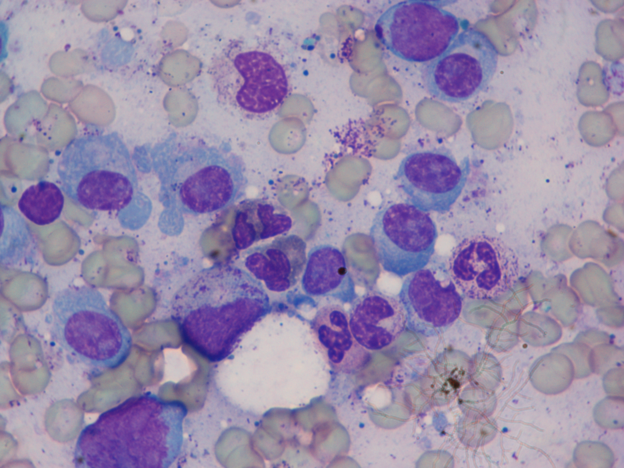
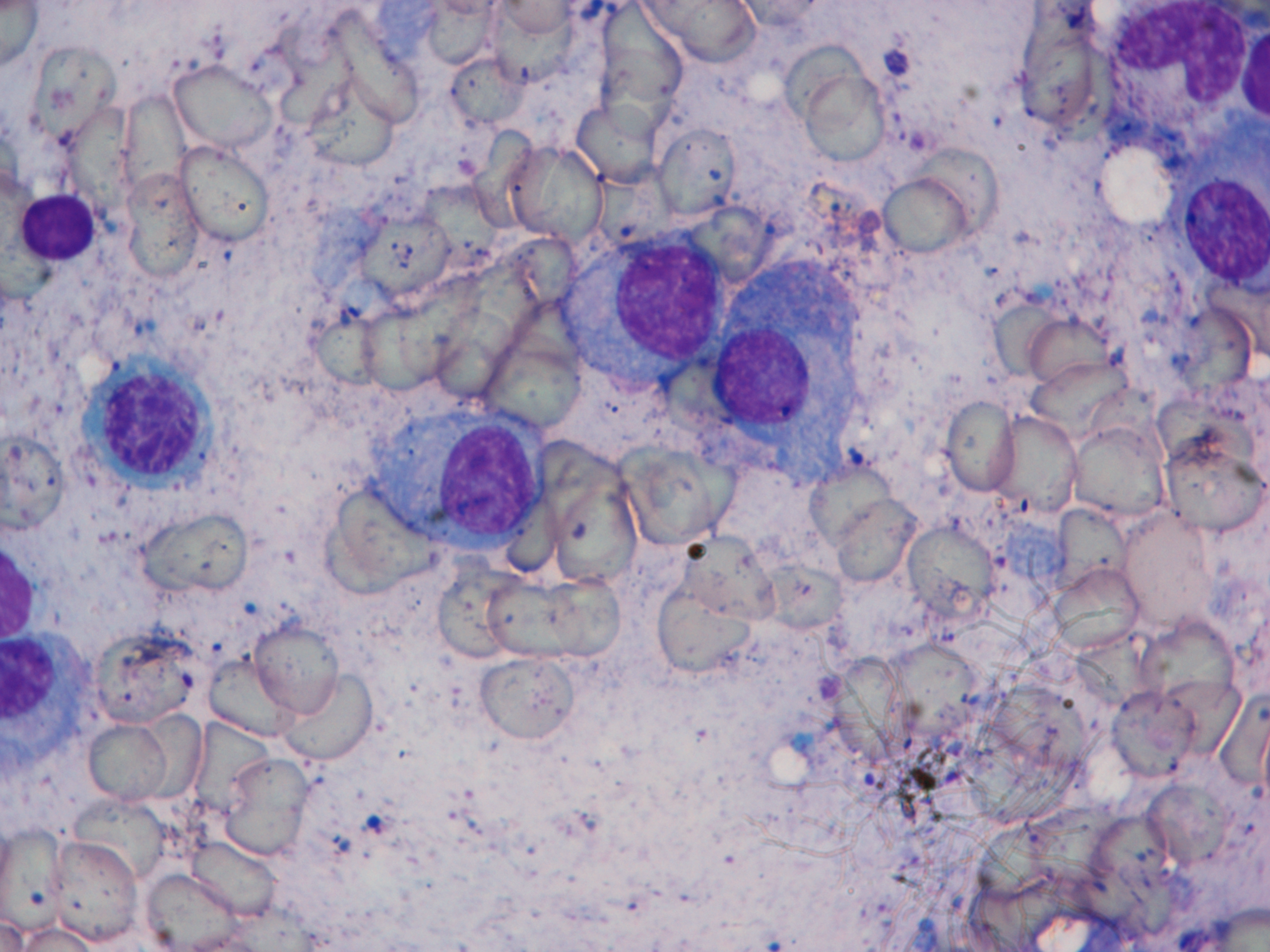
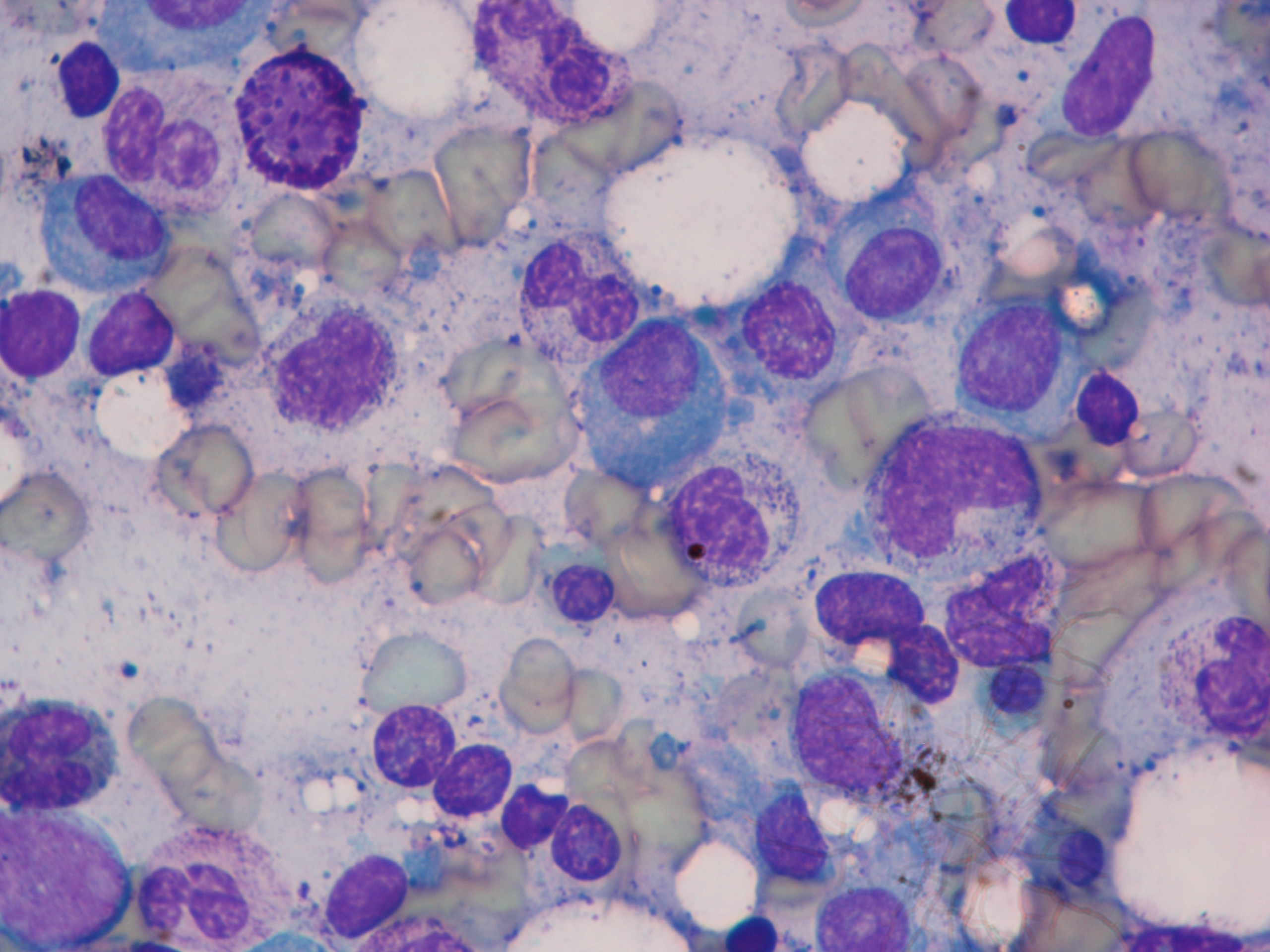
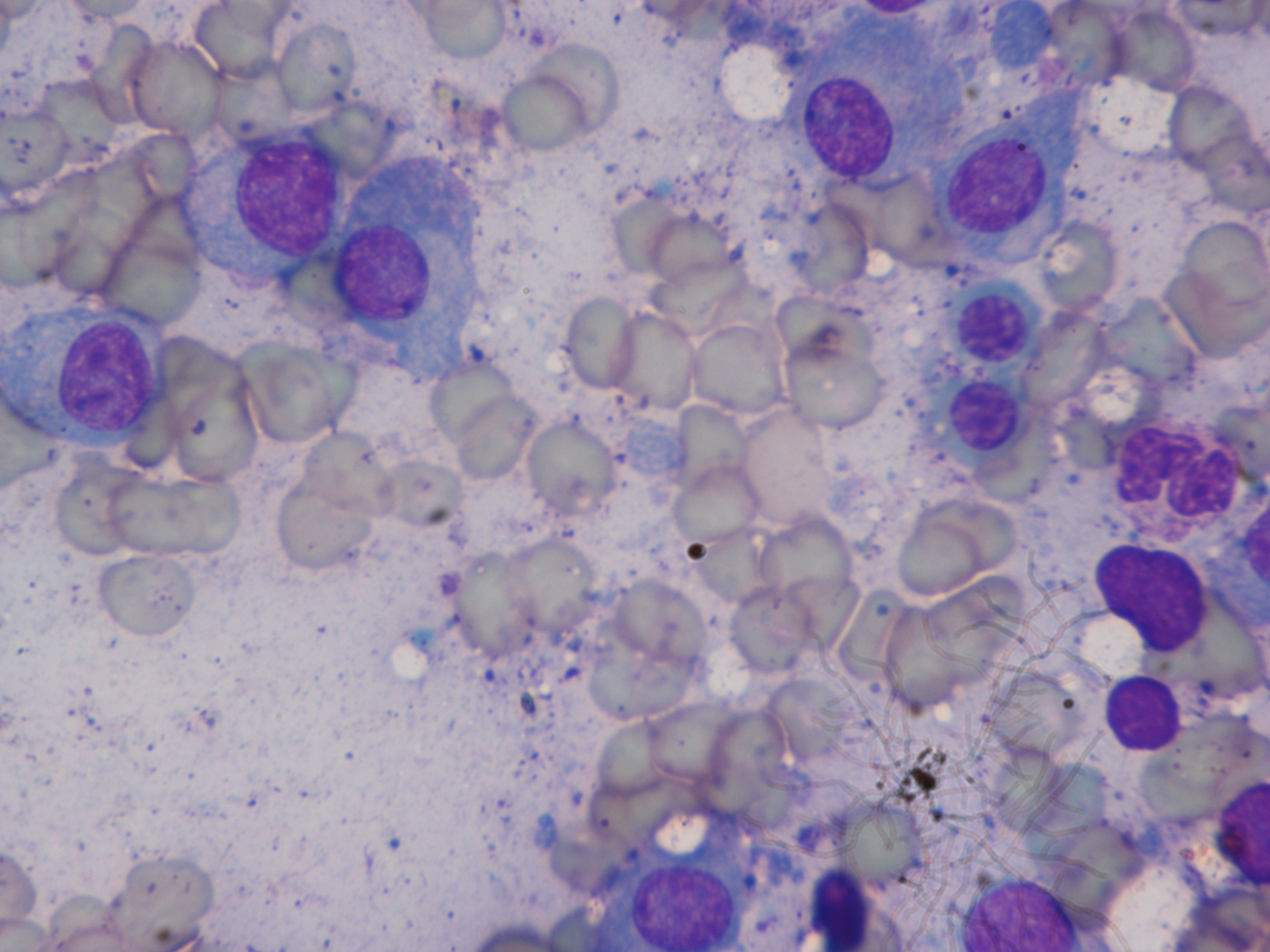
| Summary: | This dataset is a collection of 85 microscopic images of Multiple Meyloma subjects with a number of plasma cells marked by the expert pathologist. We proposed a method of color-driven multiphase levelset for automating cell segmentation in Multiple Myeloma that has been published in PLOS One Journal. The users of this dataset would be required to cite that paper. This is an effort towards building an automated pipeline for cancer detection in Multiple Myeloma. Interested researchers can propose deep learning based or advanced machine learning based solutions for plasma cell segmentation using this dataset. |
| Keywords: | Plasma cell segmentation; Multiple myeloma (MM); Blood cancer; Computer aided diagnosis; Microscopic image; Segmentation; Cancer dataset; Deep learning |
| Additional / Any Other Information: | Download |
| Release Date: | Nov. 14, 2025 |
| Access Licence Type: | Open Access |
Table 1. The sample types registered under this study are as follows:
Table 2. The samples registered under this study are as follows:
| Sample Type ID | Organism | Taxon ID | Biological Entity | Laterality | Source Tissue | Source Cell/Cell-line | Cell Organelle |
|---|---|---|---|---|---|---|---|
| HISTOSMT_10000000063 | Homo sapiens | 9606 | Hip bone | Not Applicable | Bone marrow | Normal and Plasma cells | N/A |
Table 2. The samples registered under this study are as follows:
| Sample Type ID | Sample ID | Method used for Sample Collection | Cell Phenotype Studied | Sample Source | ICD-11 Code (patient health condition) | Image category/label | Subject type |
|---|---|---|---|---|---|---|---|
| HISTOSMT_10000000063 | HISTOSM_10000297552 | Bone marrow aspiration | N/A | Laboratory Oncology Unit, Dr. B. R.A. IRCH, All India Institute of Medical Sciences (AIIMS), New Delhi, India | XH4XA9 | Stain Normalized Plasma cells | Multiple Myeloma |
| HISTOSMT_10000000063 | HISTOSM_10000297553 | Bone marrow aspiration | N/A | Laboratory Oncology Unit, Dr. B. R.A. IRCH, All India Institute of Medical Sciences (AIIMS), New Delhi, India | XH4XA9 | Stain Normalized Plasma cells | Multiple Myeloma |
| HISTOSMT_10000000063 | HISTOSM_10000297554 | Bone marrow aspiration | N/A | Laboratory Oncology Unit, Dr. B. R.A. IRCH, All India Institute of Medical Sciences (AIIMS), New Delhi, India | XH4XA9 | Stain Normalized Plasma cells | Multiple Myeloma |
| HISTOSMT_10000000063 | HISTOSM_10000297555 | Bone marrow aspiration | N/A | Laboratory Oncology Unit, Dr. B. R.A. IRCH, All India Institute of Medical Sciences (AIIMS), New Delhi, India | XH4XA9 | Stain Normalized Plasma cells | Multiple Myeloma |
| HISTOSMT_10000000063 | HISTOSM_10000297556 | Bone marrow aspiration | N/A | Laboratory Oncology Unit, Dr. B. R.A. IRCH, All India Institute of Medical Sciences (AIIMS), New Delhi, India | XH4XA9 | Stain Normalized Plasma cells | Multiple Myeloma |
| HISTOSMT_10000000063 | HISTOSM_10000297557 | Bone marrow aspiration | N/A | Laboratory Oncology Unit, Dr. B. R.A. IRCH, All India Institute of Medical Sciences (AIIMS), New Delhi, India | XH4XA9 | Stain Normalized Plasma cells | Multiple Myeloma |
| HISTOSMT_10000000063 | HISTOSM_10000297559 | Bone marrow aspiration | N/A | Laboratory Oncology Unit, Dr. B. R.A. IRCH, All India Institute of Medical Sciences (AIIMS), New Delhi, India | XH4XA9 | Stain Normalized Plasma cells | Multiple Myeloma |
| HISTOSMT_10000000063 | HISTOSM_10000297560 | Bone marrow aspiration | N/A | Laboratory Oncology Unit, Dr. B. R.A. IRCH, All India Institute of Medical Sciences (AIIMS), New Delhi, India | XH4XA9 | Stain Normalized Plasma cells | Multiple Myeloma |
| HISTOSMT_10000000063 | HISTOSM_10000297561 | Bone marrow aspiration | N/A | Laboratory Oncology Unit, Dr. B. R.A. IRCH, All India Institute of Medical Sciences (AIIMS), New Delhi, India | XH4XA9 | Stain Normalized Plasma cells | Multiple Myeloma |
| HISTOSMT_10000000063 | HISTOSM_10000297562 | Bone marrow aspiration | N/A | Laboratory Oncology Unit, Dr. B. R.A. IRCH, All India Institute of Medical Sciences (AIIMS), New Delhi, India | XH4XA9 | Stain Normalized Plasma cells | Multiple Myeloma |
| HISTOSMT_10000000063 | HISTOSM_10000297563 | Bone marrow aspiration | N/A | Laboratory Oncology Unit, Dr. B. R.A. IRCH, All India Institute of Medical Sciences (AIIMS), New Delhi, India | XH4XA9 | Stain Normalized Plasma cells | Multiple Myeloma |
| HISTOSMT_10000000063 | HISTOSM_10000297564 | Bone marrow aspiration | N/A | Laboratory Oncology Unit, Dr. B. R.A. IRCH, All India Institute of Medical Sciences (AIIMS), New Delhi, India | XH4XA9 | Stain Normalized Plasma cells | Multiple Myeloma |
| HISTOSMT_10000000063 | HISTOSM_10000297565 | Bone marrow aspiration | N/A | Laboratory Oncology Unit, Dr. B. R.A. IRCH, All India Institute of Medical Sciences (AIIMS), New Delhi, India | XH4XA9 | Stain Normalized Plasma cells | Multiple Myeloma |
| HISTOSMT_10000000063 | HISTOSM_10000297566 | Bone marrow aspiration | N/A | Laboratory Oncology Unit, Dr. B. R.A. IRCH, All India Institute of Medical Sciences (AIIMS), New Delhi, India | XH4XA9 | Stain Normalized Plasma cells | Multiple Myeloma |
| HISTOSMT_10000000063 | HISTOSM_10000297567 | Bone marrow aspiration | N/A | Laboratory Oncology Unit, Dr. B. R.A. IRCH, All India Institute of Medical Sciences (AIIMS), New Delhi, India | XH4XA9 | Stain Normalized Plasma cells | Multiple Myeloma |
| HISTOSMT_10000000063 | HISTOSM_10000297568 | Bone marrow aspiration | N/A | Laboratory Oncology Unit, Dr. B. R.A. IRCH, All India Institute of Medical Sciences (AIIMS), New Delhi, India | XH4XA9 | Stain Normalized Plasma cells | Multiple Myeloma |
| HISTOSMT_10000000063 | HISTOSM_10000297569 | Bone marrow aspiration | N/A | Laboratory Oncology Unit, Dr. B. R.A. IRCH, All India Institute of Medical Sciences (AIIMS), New Delhi, India | XH4XA9 | Stain Normalized Plasma cells | Multiple Myeloma |
| HISTOSMT_10000000063 | HISTOSM_10000297570 | Bone marrow aspiration | N/A | Laboratory Oncology Unit, Dr. B. R.A. IRCH, All India Institute of Medical Sciences (AIIMS), New Delhi, India | XH4XA9 | Stain Normalized Plasma cells | Multiple Myeloma |
| HISTOSMT_10000000063 | HISTOSM_10000297571 | Bone marrow aspiration | N/A | Laboratory Oncology Unit, Dr. B. R.A. IRCH, All India Institute of Medical Sciences (AIIMS), New Delhi, India | XH4XA9 | Stain Normalized Plasma cells | Multiple Myeloma |
| HISTOSMT_10000000063 | HISTOSM_10000297572 | Bone marrow aspiration | N/A | Laboratory Oncology Unit, Dr. B. R.A. IRCH, All India Institute of Medical Sciences (AIIMS), New Delhi, India | XH4XA9 | Stain Normalized Plasma cells | Multiple Myeloma |
| HISTOSMT_10000000063 | HISTOSM_10000297573 | Bone marrow aspiration | N/A | Laboratory Oncology Unit, Dr. B. R.A. IRCH, All India Institute of Medical Sciences (AIIMS), New Delhi, India | XH4XA9 | Stain Normalized Plasma cells | Multiple Myeloma |
| HISTOSMT_10000000063 | HISTOSM_10000297574 | Bone marrow aspiration | N/A | Laboratory Oncology Unit, Dr. B. R.A. IRCH, All India Institute of Medical Sciences (AIIMS), New Delhi, India | XH4XA9 | Stain Normalized Plasma cells | Multiple Myeloma |
| HISTOSMT_10000000063 | HISTOSM_10000297575 | Bone marrow aspiration | N/A | Laboratory Oncology Unit, Dr. B. R.A. IRCH, All India Institute of Medical Sciences (AIIMS), New Delhi, India | XH4XA9 | Stain Normalized Plasma cells | Multiple Myeloma |
| HISTOSMT_10000000063 | HISTOSM_10000297576 | Bone marrow aspiration | N/A | Laboratory Oncology Unit, Dr. B. R.A. IRCH, All India Institute of Medical Sciences (AIIMS), New Delhi, India | XH4XA9 | Stain Normalized Plasma cells | Multiple Myeloma |
| HISTOSMT_10000000063 | HISTOSM_10000297577 | Bone marrow aspiration | N/A | Laboratory Oncology Unit, Dr. B. R.A. IRCH, All India Institute of Medical Sciences (AIIMS), New Delhi, India | XH4XA9 | Stain Normalized Plasma cells | Multiple Myeloma |
| HISTOSMT_10000000063 | HISTOSM_10000297579 | Bone marrow aspiration | N/A | Laboratory Oncology Unit, Dr. B. R.A. IRCH, All India Institute of Medical Sciences (AIIMS), New Delhi, India | XH4XA9 | Stain Normalized Plasma cells | Multiple Myeloma |
| HISTOSMT_10000000063 | HISTOSM_10000297580 | Bone marrow aspiration | N/A | Laboratory Oncology Unit, Dr. B. R.A. IRCH, All India Institute of Medical Sciences (AIIMS), New Delhi, India | XH4XA9 | Stain Normalized Plasma cells | Multiple Myeloma |
| HISTOSMT_10000000063 | HISTOSM_10000297581 | Bone marrow aspiration | N/A | Laboratory Oncology Unit, Dr. B. R.A. IRCH, All India Institute of Medical Sciences (AIIMS), New Delhi, India | XH4XA9 | Stain Normalized Plasma cells | Multiple Myeloma |
| HISTOSMT_10000000063 | HISTOSM_10000297582 | Bone marrow aspiration | N/A | Laboratory Oncology Unit, Dr. B. R.A. IRCH, All India Institute of Medical Sciences (AIIMS), New Delhi, India | XH4XA9 | Stain Normalized Plasma cells | Multiple Myeloma |
| HISTOSMT_10000000063 | HISTOSM_10000297583 | Bone marrow aspiration | N/A | Laboratory Oncology Unit, Dr. B. R.A. IRCH, All India Institute of Medical Sciences (AIIMS), New Delhi, India | XH4XA9 | Stain Normalized Plasma cells | Multiple Myeloma |
| HISTOSMT_10000000063 | HISTOSM_10000297584 | Bone marrow aspiration | N/A | Laboratory Oncology Unit, Dr. B. R.A. IRCH, All India Institute of Medical Sciences (AIIMS), New Delhi, India | XH4XA9 | Stain Normalized Plasma cells | Multiple Myeloma |
| HISTOSMT_10000000063 | HISTOSM_10000297585 | Bone marrow aspiration | N/A | Laboratory Oncology Unit, Dr. B. R.A. IRCH, All India Institute of Medical Sciences (AIIMS), New Delhi, India | XH4XA9 | Stain Normalized Plasma cells | Multiple Myeloma |
| HISTOSMT_10000000063 | HISTOSM_10000297586 | Bone marrow aspiration | N/A | Laboratory Oncology Unit, Dr. B. R.A. IRCH, All India Institute of Medical Sciences (AIIMS), New Delhi, India | XH4XA9 | Stain Normalized Plasma cells | Multiple Myeloma |
| HISTOSMT_10000000063 | HISTOSM_10000297587 | Bone marrow aspiration | N/A | Laboratory Oncology Unit, Dr. B. R.A. IRCH, All India Institute of Medical Sciences (AIIMS), New Delhi, India | XH4XA9 | Stain Normalized Plasma cells | Multiple Myeloma |
| HISTOSMT_10000000063 | HISTOSM_10000297588 | Bone marrow aspiration | N/A | Laboratory Oncology Unit, Dr. B. R.A. IRCH, All India Institute of Medical Sciences (AIIMS), New Delhi, India | XH4XA9 | Stain Normalized Plasma cells | Multiple Myeloma |
| HISTOSMT_10000000063 | HISTOSM_10000297589 | Bone marrow aspiration | N/A | Laboratory Oncology Unit, Dr. B. R.A. IRCH, All India Institute of Medical Sciences (AIIMS), New Delhi, India | XH4XA9 | Stain Normalized Plasma cells | Multiple Myeloma |
| HISTOSMT_10000000063 | HISTOSM_10000297590 | Bone marrow aspiration | N/A | Laboratory Oncology Unit, Dr. B. R.A. IRCH, All India Institute of Medical Sciences (AIIMS), New Delhi, India | XH4XA9 | Stain Normalized Plasma cells | Multiple Myeloma |
| HISTOSMT_10000000063 | HISTOSM_10000297591 | Bone marrow aspiration | N/A | Laboratory Oncology Unit, Dr. B. R.A. IRCH, All India Institute of Medical Sciences (AIIMS), New Delhi, India | XH4XA9 | Stain Normalized Plasma cells | Multiple Myeloma |
| HISTOSMT_10000000063 | HISTOSM_10000297592 | Bone marrow aspiration | N/A | Laboratory Oncology Unit, Dr. B. R.A. IRCH, All India Institute of Medical Sciences (AIIMS), New Delhi, India | XH4XA9 | Stain Normalized Plasma cells | Multiple Myeloma |
| HISTOSMT_10000000063 | HISTOSM_10000297593 | Bone marrow aspiration | N/A | Laboratory Oncology Unit, Dr. B. R.A. IRCH, All India Institute of Medical Sciences (AIIMS), New Delhi, India | XH4XA9 | Stain Normalized Plasma cells | Multiple Myeloma |
| HISTOSMT_10000000063 | HISTOSM_10000297594 | Bone marrow aspiration | N/A | Laboratory Oncology Unit, Dr. B. R.A. IRCH, All India Institute of Medical Sciences (AIIMS), New Delhi, India | XH4XA9 | Stain Normalized Plasma cells | Multiple Myeloma |
| HISTOSMT_10000000063 | HISTOSM_10000297595 | Bone marrow aspiration | N/A | Laboratory Oncology Unit, Dr. B. R.A. IRCH, All India Institute of Medical Sciences (AIIMS), New Delhi, India | XH4XA9 | Stain Normalized Plasma cells | Multiple Myeloma |
| HISTOSMT_10000000063 | HISTOSM_10000297596 | Bone marrow aspiration | N/A | Laboratory Oncology Unit, Dr. B. R.A. IRCH, All India Institute of Medical Sciences (AIIMS), New Delhi, India | XH4XA9 | Stain Normalized Plasma cells | Multiple Myeloma |
| HISTOSMT_10000000063 | HISTOSM_10000297597 | Bone marrow aspiration | N/A | Laboratory Oncology Unit, Dr. B. R.A. IRCH, All India Institute of Medical Sciences (AIIMS), New Delhi, India | XH4XA9 | Stain Normalized Plasma cells | Multiple Myeloma |
| HISTOSMT_10000000063 | HISTOSM_10000297599 | Bone marrow aspiration | N/A | Laboratory Oncology Unit, Dr. B. R.A. IRCH, All India Institute of Medical Sciences (AIIMS), New Delhi, India | XH4XA9 | Stain Normalized Plasma cells | Multiple Myeloma |
| HISTOSMT_10000000063 | HISTOSM_10000297600 | Bone marrow aspiration | N/A | Laboratory Oncology Unit, Dr. B. R.A. IRCH, All India Institute of Medical Sciences (AIIMS), New Delhi, India | XH4XA9 | Stain Normalized Plasma cells | Multiple Myeloma |
| HISTOSMT_10000000063 | HISTOSM_10000297601 | Bone marrow aspiration | N/A | Laboratory Oncology Unit, Dr. B. R.A. IRCH, All India Institute of Medical Sciences (AIIMS), New Delhi, India | XH4XA9 | Stain Normalized Plasma cells | Multiple Myeloma |
| HISTOSMT_10000000063 | HISTOSM_10000297602 | Bone marrow aspiration | N/A | Laboratory Oncology Unit, Dr. B. R.A. IRCH, All India Institute of Medical Sciences (AIIMS), New Delhi, India | XH4XA9 | Stain Normalized Plasma cells | Multiple Myeloma |
| HISTOSMT_10000000063 | HISTOSM_10000297603 | Bone marrow aspiration | N/A | Laboratory Oncology Unit, Dr. B. R.A. IRCH, All India Institute of Medical Sciences (AIIMS), New Delhi, India | XH4XA9 | Stain Normalized Plasma cells | Multiple Myeloma |
| HISTOSMT_10000000063 | HISTOSM_10000297604 | Bone marrow aspiration | N/A | Laboratory Oncology Unit, Dr. B. R.A. IRCH, All India Institute of Medical Sciences (AIIMS), New Delhi, India | XH4XA9 | Stain Normalized Plasma cells | Multiple Myeloma |
| HISTOSMT_10000000063 | HISTOSM_10000297605 | Bone marrow aspiration | N/A | Laboratory Oncology Unit, Dr. B. R.A. IRCH, All India Institute of Medical Sciences (AIIMS), New Delhi, India | XH4XA9 | Stain Normalized Plasma cells | Multiple Myeloma |
| HISTOSMT_10000000063 | HISTOSM_10000297606 | Bone marrow aspiration | N/A | Laboratory Oncology Unit, Dr. B. R.A. IRCH, All India Institute of Medical Sciences (AIIMS), New Delhi, India | XH4XA9 | Stain Normalized Plasma cells | Multiple Myeloma |
| HISTOSMT_10000000063 | HISTOSM_10000297607 | Bone marrow aspiration | N/A | Laboratory Oncology Unit, Dr. B. R.A. IRCH, All India Institute of Medical Sciences (AIIMS), New Delhi, India | XH4XA9 | Stain Normalized Plasma cells | Multiple Myeloma |
| HISTOSMT_10000000063 | HISTOSM_10000297608 | Bone marrow aspiration | N/A | Laboratory Oncology Unit, Dr. B. R.A. IRCH, All India Institute of Medical Sciences (AIIMS), New Delhi, India | XH4XA9 | Stain Normalized Plasma cells | Multiple Myeloma |
| HISTOSMT_10000000063 | HISTOSM_10000297609 | Bone marrow aspiration | N/A | Laboratory Oncology Unit, Dr. B. R.A. IRCH, All India Institute of Medical Sciences (AIIMS), New Delhi, India | XH4XA9 | Stain Normalized Plasma cells | Multiple Myeloma |
| HISTOSMT_10000000063 | HISTOSM_10000297610 | Bone marrow aspiration | N/A | Laboratory Oncology Unit, Dr. B. R.A. IRCH, All India Institute of Medical Sciences (AIIMS), New Delhi, India | XH4XA9 | Stain Normalized Plasma cells | Multiple Myeloma |
| HISTOSMT_10000000063 | HISTOSM_10000297611 | Bone marrow aspiration | N/A | Laboratory Oncology Unit, Dr. B. R.A. IRCH, All India Institute of Medical Sciences (AIIMS), New Delhi, India | XH4XA9 | Stain Normalized Plasma cells | Multiple Myeloma |
| HISTOSMT_10000000063 | HISTOSM_10000297612 | Bone marrow aspiration | N/A | Laboratory Oncology Unit, Dr. B. R.A. IRCH, All India Institute of Medical Sciences (AIIMS), New Delhi, India | XH4XA9 | Stain Normalized Plasma cells | Multiple Myeloma |
| HISTOSMT_10000000063 | HISTOSM_10000297613 | Bone marrow aspiration | N/A | Laboratory Oncology Unit, Dr. B. R.A. IRCH, All India Institute of Medical Sciences (AIIMS), New Delhi, India | XH4XA9 | Stain Normalized Plasma cells | Multiple Myeloma |
| HISTOSMT_10000000063 | HISTOSM_10000297614 | Bone marrow aspiration | N/A | Laboratory Oncology Unit, Dr. B. R.A. IRCH, All India Institute of Medical Sciences (AIIMS), New Delhi, India | XH4XA9 | Stain Normalized Plasma cells | Multiple Myeloma |
| HISTOSMT_10000000063 | HISTOSM_10000297615 | Bone marrow aspiration | N/A | Laboratory Oncology Unit, Dr. B. R.A. IRCH, All India Institute of Medical Sciences (AIIMS), New Delhi, India | XH4XA9 | Stain Normalized Plasma cells | Multiple Myeloma |
| HISTOSMT_10000000063 | HISTOSM_10000297616 | Bone marrow aspiration | N/A | Laboratory Oncology Unit, Dr. B. R.A. IRCH, All India Institute of Medical Sciences (AIIMS), New Delhi, India | XH4XA9 | Stain Normalized Plasma cells | Multiple Myeloma |
| HISTOSMT_10000000063 | HISTOSM_10000297617 | Bone marrow aspiration | N/A | Laboratory Oncology Unit, Dr. B. R.A. IRCH, All India Institute of Medical Sciences (AIIMS), New Delhi, India | XH4XA9 | Stain Normalized Plasma cells | Multiple Myeloma |
| HISTOSMT_10000000063 | HISTOSM_10000297551 | Bone marrow aspiration | N/A | Laboratory Oncology Unit, Dr. B. R.A. IRCH, All India Institute of Medical Sciences (AIIMS), New Delhi, India | XH4XA9 | Stain Normalized Plasma cells | Multiple Myeloma |
| HISTOSMT_10000000063 | HISTOSM_10000297558 | Bone marrow aspiration | N/A | Laboratory Oncology Unit, Dr. B. R.A. IRCH, All India Institute of Medical Sciences (AIIMS), New Delhi, India | XH4XA9 | Stain Normalized Plasma cells | Multiple Myeloma |
| HISTOSMT_10000000063 | HISTOSM_10000297619 | Bone marrow aspiration | N/A | Laboratory Oncology Unit, Dr. B. R.A. IRCH, All India Institute of Medical Sciences (AIIMS), New Delhi, India | XH4XA9 | Stain Normalized Plasma cells | Multiple Myeloma |
| HISTOSMT_10000000063 | HISTOSM_10000297620 | Bone marrow aspiration | N/A | Laboratory Oncology Unit, Dr. B. R.A. IRCH, All India Institute of Medical Sciences (AIIMS), New Delhi, India | XH4XA9 | Stain Normalized Plasma cells | Multiple Myeloma |
| HISTOSMT_10000000063 | HISTOSM_10000297621 | Bone marrow aspiration | N/A | Laboratory Oncology Unit, Dr. B. R.A. IRCH, All India Institute of Medical Sciences (AIIMS), New Delhi, India | XH4XA9 | Stain Normalized Plasma cells | Multiple Myeloma |
| HISTOSMT_10000000063 | HISTOSM_10000297622 | Bone marrow aspiration | N/A | Laboratory Oncology Unit, Dr. B. R.A. IRCH, All India Institute of Medical Sciences (AIIMS), New Delhi, India | XH4XA9 | Stain Normalized Plasma cells | Multiple Myeloma |
| HISTOSMT_10000000063 | HISTOSM_10000297623 | Bone marrow aspiration | N/A | Laboratory Oncology Unit, Dr. B. R.A. IRCH, All India Institute of Medical Sciences (AIIMS), New Delhi, India | XH4XA9 | Stain Normalized Plasma cells | Multiple Myeloma |
| HISTOSMT_10000000063 | HISTOSM_10000297624 | Bone marrow aspiration | N/A | Laboratory Oncology Unit, Dr. B. R.A. IRCH, All India Institute of Medical Sciences (AIIMS), New Delhi, India | XH4XA9 | Stain Normalized Plasma cells | Multiple Myeloma |
| HISTOSMT_10000000063 | HISTOSM_10000297625 | Bone marrow aspiration | N/A | Laboratory Oncology Unit, Dr. B. R.A. IRCH, All India Institute of Medical Sciences (AIIMS), New Delhi, India | XH4XA9 | Stain Normalized Plasma cells | Multiple Myeloma |
| HISTOSMT_10000000063 | HISTOSM_10000297626 | Bone marrow aspiration | N/A | Laboratory Oncology Unit, Dr. B. R.A. IRCH, All India Institute of Medical Sciences (AIIMS), New Delhi, India | XH4XA9 | Stain Normalized Plasma cells | Multiple Myeloma |
| HISTOSMT_10000000063 | HISTOSM_10000297627 | Bone marrow aspiration | N/A | Laboratory Oncology Unit, Dr. B. R.A. IRCH, All India Institute of Medical Sciences (AIIMS), New Delhi, India | XH4XA9 | Stain Normalized Plasma cells | Multiple Myeloma |
| HISTOSMT_10000000063 | HISTOSM_10000297628 | Bone marrow aspiration | N/A | Laboratory Oncology Unit, Dr. B. R.A. IRCH, All India Institute of Medical Sciences (AIIMS), New Delhi, India | XH4XA9 | Stain Normalized Plasma cells | Multiple Myeloma |
| HISTOSMT_10000000063 | HISTOSM_10000297629 | Bone marrow aspiration | N/A | Laboratory Oncology Unit, Dr. B. R.A. IRCH, All India Institute of Medical Sciences (AIIMS), New Delhi, India | XH4XA9 | Stain Normalized Plasma cells | Multiple Myeloma |
| HISTOSMT_10000000063 | HISTOSM_10000297630 | Bone marrow aspiration | N/A | Laboratory Oncology Unit, Dr. B. R.A. IRCH, All India Institute of Medical Sciences (AIIMS), New Delhi, India | XH4XA9 | Stain Normalized Plasma cells | Multiple Myeloma |
| HISTOSMT_10000000063 | HISTOSM_10000297631 | Bone marrow aspiration | N/A | Laboratory Oncology Unit, Dr. B. R.A. IRCH, All India Institute of Medical Sciences (AIIMS), New Delhi, India | XH4XA9 | Stain Normalized Plasma cells | Multiple Myeloma |
| HISTOSMT_10000000063 | HISTOSM_10000297632 | Bone marrow aspiration | N/A | Laboratory Oncology Unit, Dr. B. R.A. IRCH, All India Institute of Medical Sciences (AIIMS), New Delhi, India | XH4XA9 | Stain Normalized Plasma cells | Multiple Myeloma |
| HISTOSMT_10000000063 | HISTOSM_10000297633 | Bone marrow aspiration | N/A | Laboratory Oncology Unit, Dr. B. R.A. IRCH, All India Institute of Medical Sciences (AIIMS), New Delhi, India | XH4XA9 | Stain Normalized Plasma cells | Multiple Myeloma |
| HISTOSMT_10000000063 | HISTOSM_10000297634 | Bone marrow aspiration | N/A | Laboratory Oncology Unit, Dr. B. R.A. IRCH, All India Institute of Medical Sciences (AIIMS), New Delhi, India | XH4XA9 | Stain Normalized Plasma cells | Multiple Myeloma |
| HISTOSMT_10000000063 | HISTOSM_10000297635 | Bone marrow aspiration | N/A | Laboratory Oncology Unit, Dr. B. R.A. IRCH, All India Institute of Medical Sciences (AIIMS), New Delhi, India | XH4XA9 | Stain Normalized Plasma cells | Multiple Myeloma |
| HISTOSMT_10000000063 | HISTOSM_10000297578 | Bone marrow aspiration | N/A | Laboratory Oncology Unit, Dr. B. R.A. IRCH, All India Institute of Medical Sciences (AIIMS), New Delhi, India | XH4XA9 | Stain Normalized Plasma cells | Multiple Myeloma |
| HISTOSMT_10000000063 | HISTOSM_10000297598 | Bone marrow aspiration | N/A | Laboratory Oncology Unit, Dr. B. R.A. IRCH, All India Institute of Medical Sciences (AIIMS), New Delhi, India | XH4XA9 | Stain Normalized Plasma cells | Multiple Myeloma |
| HISTOSMT_10000000063 | HISTOSM_10000297618 | Bone marrow aspiration | N/A | Laboratory Oncology Unit, Dr. B. R.A. IRCH, All India Institute of Medical Sciences (AIIMS), New Delhi, India | XH4XA9 | Stain Normalized Plasma cells | Multiple Myeloma |
Table 3. The experiment types registered under this study are as follows:
| Experiment Type ID | Instrument Name | Instrument Type | Manufacturer | Model |
|---|---|---|---|---|
| HISTOET_10000000029 | Camera | Microscope | Nikon | Eclipse-200 |
| Experimental Design Summary (HISTOET_10000000029) |
|---|
| Microscopic images were captured from bone marrow aspirate slides of patients diagnosed with multiple myeloma as per the standard guidelines. Slides were stained using Jenner-Giemsa stain. Images were captured at 1000x magnification using Nikon Eclipse-200 microscope equipped with a digital camera. Images were captured in raw BMP format with a size of 2560x1920 pixels. In all, this dataset consists of 85 images. All these 85 images were stain normalized using our in-house methodology before being used for segmentation. These stain normalized images have been provided as the annotated dataset with plasma cells marked in all image slides contained in a presentation for the ready reference of readers. Please refer to the “Additional File”, at the “Project” section of IBIA for more details. |
| Acquired Images Annotation Description (HISTOET_10000000029) |
|---|
| The plasma cells in the microscopic images of Multiple Meyloma subjects were marked by the expert pathologist. |
Table 4. The experiments registered under this study are as follows:
| Sample ID | Experiment Type ID | Experiment ID | Image type (Original / Derived / Unknown) | Any Other Information | Staining Type | Images Magnification | Tissue / Tumor Fixative Used | Camera Used to Capture Images | Data Repository Name (If already deposited in another repository) | Licence Type (original source) | Stain Normalization Method |
|---|---|---|---|---|---|---|---|---|---|---|---|
| HISTOSM_10000297551 | HISTOET_10000000029 | HISTOE_10000269965 | Derived | N/A | Jenner-Giemsa | 1000x | N/A | Nikon Eclipse-200 | The Cancer Imaging Archive (TCIA) and Harvard Dataverse | CC BY 3.0 | in-house methodology |
| HISTOSM_10000297552 | HISTOET_10000000029 | HISTOE_10000269966 | Derived | N/A | Jenner-Giemsa | 1000x | N/A | Nikon Eclipse-200 | The Cancer Imaging Archive (TCIA) and Harvard Dataverse | CC BY 3.0 | in-house methodology |
| HISTOSM_10000297553 | HISTOET_10000000029 | HISTOE_10000269967 | Derived | N/A | Jenner-Giemsa | 1000x | N/A | Nikon Eclipse-200 | The Cancer Imaging Archive (TCIA) and Harvard Dataverse | CC BY 3.0 | in-house methodology |
| HISTOSM_10000297554 | HISTOET_10000000029 | HISTOE_10000269968 | Derived | N/A | Jenner-Giemsa | 1000x | N/A | Nikon Eclipse-200 | The Cancer Imaging Archive (TCIA) and Harvard Dataverse | CC BY 3.0 | in-house methodology |
| HISTOSM_10000297555 | HISTOET_10000000029 | HISTOE_10000269969 | Derived | N/A | Jenner-Giemsa | 1000x | N/A | Nikon Eclipse-200 | The Cancer Imaging Archive (TCIA) and Harvard Dataverse | CC BY 3.0 | in-house methodology |
| HISTOSM_10000297556 | HISTOET_10000000029 | HISTOE_10000269970 | Derived | N/A | Jenner-Giemsa | 1000x | N/A | Nikon Eclipse-200 | The Cancer Imaging Archive (TCIA) and Harvard Dataverse | CC BY 3.0 | in-house methodology |
| HISTOSM_10000297557 | HISTOET_10000000029 | HISTOE_10000269971 | Derived | N/A | Jenner-Giemsa | 1000x | N/A | Nikon Eclipse-200 | The Cancer Imaging Archive (TCIA) and Harvard Dataverse | CC BY 3.0 | in-house methodology |
| HISTOSM_10000297558 | HISTOET_10000000029 | HISTOE_10000269972 | Derived | N/A | Jenner-Giemsa | 1000x | N/A | Nikon Eclipse-200 | The Cancer Imaging Archive (TCIA) and Harvard Dataverse | CC BY 3.0 | in-house methodology |
| HISTOSM_10000297559 | HISTOET_10000000029 | HISTOE_10000269973 | Derived | N/A | Jenner-Giemsa | 1000x | N/A | Nikon Eclipse-200 | The Cancer Imaging Archive (TCIA) and Harvard Dataverse | CC BY 3.0 | in-house methodology |
| HISTOSM_10000297560 | HISTOET_10000000029 | HISTOE_10000269974 | Derived | N/A | Jenner-Giemsa | 1000x | N/A | Nikon Eclipse-200 | The Cancer Imaging Archive (TCIA) and Harvard Dataverse | CC BY 3.0 | in-house methodology |
| HISTOSM_10000297561 | HISTOET_10000000029 | HISTOE_10000269975 | Derived | N/A | Jenner-Giemsa | 1000x | N/A | Nikon Eclipse-200 | The Cancer Imaging Archive (TCIA) and Harvard Dataverse | CC BY 3.0 | in-house methodology |
| HISTOSM_10000297562 | HISTOET_10000000029 | HISTOE_10000269976 | Derived | N/A | Jenner-Giemsa | 1000x | N/A | Nikon Eclipse-200 | The Cancer Imaging Archive (TCIA) and Harvard Dataverse | CC BY 3.0 | in-house methodology |
| HISTOSM_10000297563 | HISTOET_10000000029 | HISTOE_10000269977 | Derived | N/A | Jenner-Giemsa | 1000x | N/A | Nikon Eclipse-200 | The Cancer Imaging Archive (TCIA) and Harvard Dataverse | CC BY 3.0 | in-house methodology |
| HISTOSM_10000297564 | HISTOET_10000000029 | HISTOE_10000269978 | Derived | N/A | Jenner-Giemsa | 1000x | N/A | Nikon Eclipse-200 | The Cancer Imaging Archive (TCIA) and Harvard Dataverse | CC BY 3.0 | in-house methodology |
| HISTOSM_10000297565 | HISTOET_10000000029 | HISTOE_10000269979 | Derived | N/A | Jenner-Giemsa | 1000x | N/A | Nikon Eclipse-200 | The Cancer Imaging Archive (TCIA) and Harvard Dataverse | CC BY 3.0 | in-house methodology |
| HISTOSM_10000297566 | HISTOET_10000000029 | HISTOE_10000269980 | Derived | N/A | Jenner-Giemsa | 1000x | N/A | Nikon Eclipse-200 | The Cancer Imaging Archive (TCIA) and Harvard Dataverse | CC BY 3.0 | in-house methodology |
| HISTOSM_10000297567 | HISTOET_10000000029 | HISTOE_10000269981 | Derived | N/A | Jenner-Giemsa | 1000x | N/A | Nikon Eclipse-200 | The Cancer Imaging Archive (TCIA) and Harvard Dataverse | CC BY 3.0 | in-house methodology |
| HISTOSM_10000297568 | HISTOET_10000000029 | HISTOE_10000269982 | Derived | N/A | Jenner-Giemsa | 1000x | N/A | Nikon Eclipse-200 | The Cancer Imaging Archive (TCIA) and Harvard Dataverse | CC BY 3.0 | in-house methodology |
| HISTOSM_10000297569 | HISTOET_10000000029 | HISTOE_10000269983 | Derived | N/A | Jenner-Giemsa | 1000x | N/A | Nikon Eclipse-200 | The Cancer Imaging Archive (TCIA) and Harvard Dataverse | CC BY 3.0 | in-house methodology |
| HISTOSM_10000297570 | HISTOET_10000000029 | HISTOE_10000269984 | Derived | N/A | Jenner-Giemsa | 1000x | N/A | Nikon Eclipse-200 | The Cancer Imaging Archive (TCIA) and Harvard Dataverse | CC BY 3.0 | in-house methodology |
| HISTOSM_10000297571 | HISTOET_10000000029 | HISTOE_10000269985 | Derived | N/A | Jenner-Giemsa | 1000x | N/A | Nikon Eclipse-200 | The Cancer Imaging Archive (TCIA) and Harvard Dataverse | CC BY 3.0 | in-house methodology |
| HISTOSM_10000297572 | HISTOET_10000000029 | HISTOE_10000269986 | Derived | N/A | Jenner-Giemsa | 1000x | N/A | Nikon Eclipse-200 | The Cancer Imaging Archive (TCIA) and Harvard Dataverse | CC BY 3.0 | in-house methodology |
| HISTOSM_10000297573 | HISTOET_10000000029 | HISTOE_10000269987 | Derived | N/A | Jenner-Giemsa | 1000x | N/A | Nikon Eclipse-200 | The Cancer Imaging Archive (TCIA) and Harvard Dataverse | CC BY 3.0 | in-house methodology |
| HISTOSM_10000297574 | HISTOET_10000000029 | HISTOE_10000269988 | Derived | N/A | Jenner-Giemsa | 1000x | N/A | Nikon Eclipse-200 | The Cancer Imaging Archive (TCIA) and Harvard Dataverse | CC BY 3.0 | in-house methodology |
| HISTOSM_10000297575 | HISTOET_10000000029 | HISTOE_10000269989 | Derived | N/A | Jenner-Giemsa | 1000x | N/A | Nikon Eclipse-200 | The Cancer Imaging Archive (TCIA) and Harvard Dataverse | CC BY 3.0 | in-house methodology |
| HISTOSM_10000297576 | HISTOET_10000000029 | HISTOE_10000269990 | Derived | N/A | Jenner-Giemsa | 1000x | N/A | Nikon Eclipse-200 | The Cancer Imaging Archive (TCIA) and Harvard Dataverse | CC BY 3.0 | in-house methodology |
| HISTOSM_10000297577 | HISTOET_10000000029 | HISTOE_10000269991 | Derived | N/A | Jenner-Giemsa | 1000x | N/A | Nikon Eclipse-200 | The Cancer Imaging Archive (TCIA) and Harvard Dataverse | CC BY 3.0 | in-house methodology |
| HISTOSM_10000297578 | HISTOET_10000000029 | HISTOE_10000269992 | Derived | N/A | Jenner-Giemsa | 1000x | N/A | Nikon Eclipse-200 | The Cancer Imaging Archive (TCIA) and Harvard Dataverse | CC BY 3.0 | in-house methodology |
| HISTOSM_10000297579 | HISTOET_10000000029 | HISTOE_10000269993 | Derived | N/A | Jenner-Giemsa | 1000x | N/A | Nikon Eclipse-200 | The Cancer Imaging Archive (TCIA) and Harvard Dataverse | CC BY 3.0 | in-house methodology |
| HISTOSM_10000297580 | HISTOET_10000000029 | HISTOE_10000269994 | Derived | N/A | Jenner-Giemsa | 1000x | N/A | Nikon Eclipse-200 | The Cancer Imaging Archive (TCIA) and Harvard Dataverse | CC BY 3.0 | in-house methodology |
| HISTOSM_10000297581 | HISTOET_10000000029 | HISTOE_10000269995 | Derived | N/A | Jenner-Giemsa | 1000x | N/A | Nikon Eclipse-200 | The Cancer Imaging Archive (TCIA) and Harvard Dataverse | CC BY 3.0 | in-house methodology |
| HISTOSM_10000297582 | HISTOET_10000000029 | HISTOE_10000269996 | Derived | N/A | Jenner-Giemsa | 1000x | N/A | Nikon Eclipse-200 | The Cancer Imaging Archive (TCIA) and Harvard Dataverse | CC BY 3.0 | in-house methodology |
| HISTOSM_10000297583 | HISTOET_10000000029 | HISTOE_10000269997 | Derived | N/A | Jenner-Giemsa | 1000x | N/A | Nikon Eclipse-200 | The Cancer Imaging Archive (TCIA) and Harvard Dataverse | CC BY 3.0 | in-house methodology |
| HISTOSM_10000297584 | HISTOET_10000000029 | HISTOE_10000269998 | Derived | N/A | Jenner-Giemsa | 1000x | N/A | Nikon Eclipse-200 | The Cancer Imaging Archive (TCIA) and Harvard Dataverse | CC BY 3.0 | in-house methodology |
| HISTOSM_10000297585 | HISTOET_10000000029 | HISTOE_10000269999 | Derived | N/A | Jenner-Giemsa | 1000x | N/A | Nikon Eclipse-200 | The Cancer Imaging Archive (TCIA) and Harvard Dataverse | CC BY 3.0 | in-house methodology |
| HISTOSM_10000297586 | HISTOET_10000000029 | HISTOE_10000270000 | Derived | N/A | Jenner-Giemsa | 1000x | N/A | Nikon Eclipse-200 | The Cancer Imaging Archive (TCIA) and Harvard Dataverse | CC BY 3.0 | in-house methodology |
| HISTOSM_10000297587 | HISTOET_10000000029 | HISTOE_10000270001 | Derived | N/A | Jenner-Giemsa | 1000x | N/A | Nikon Eclipse-200 | The Cancer Imaging Archive (TCIA) and Harvard Dataverse | CC BY 3.0 | in-house methodology |
| HISTOSM_10000297588 | HISTOET_10000000029 | HISTOE_10000270002 | Derived | N/A | Jenner-Giemsa | 1000x | N/A | Nikon Eclipse-200 | The Cancer Imaging Archive (TCIA) and Harvard Dataverse | CC BY 3.0 | in-house methodology |
| HISTOSM_10000297589 | HISTOET_10000000029 | HISTOE_10000270003 | Derived | N/A | Jenner-Giemsa | 1000x | N/A | Nikon Eclipse-200 | The Cancer Imaging Archive (TCIA) and Harvard Dataverse | CC BY 3.0 | in-house methodology |
| HISTOSM_10000297590 | HISTOET_10000000029 | HISTOE_10000270004 | Derived | N/A | Jenner-Giemsa | 1000x | N/A | Nikon Eclipse-200 | The Cancer Imaging Archive (TCIA) and Harvard Dataverse | CC BY 3.0 | in-house methodology |
| HISTOSM_10000297591 | HISTOET_10000000029 | HISTOE_10000270005 | Derived | N/A | Jenner-Giemsa | 1000x | N/A | Nikon Eclipse-200 | The Cancer Imaging Archive (TCIA) and Harvard Dataverse | CC BY 3.0 | in-house methodology |
| HISTOSM_10000297592 | HISTOET_10000000029 | HISTOE_10000270006 | Derived | N/A | Jenner-Giemsa | 1000x | N/A | Nikon Eclipse-200 | The Cancer Imaging Archive (TCIA) and Harvard Dataverse | CC BY 3.0 | in-house methodology |
| HISTOSM_10000297593 | HISTOET_10000000029 | HISTOE_10000270007 | Derived | N/A | Jenner-Giemsa | 1000x | N/A | Nikon Eclipse-200 | The Cancer Imaging Archive (TCIA) and Harvard Dataverse | CC BY 3.0 | in-house methodology |
| HISTOSM_10000297594 | HISTOET_10000000029 | HISTOE_10000270008 | Derived | N/A | Jenner-Giemsa | 1000x | N/A | Nikon Eclipse-200 | The Cancer Imaging Archive (TCIA) and Harvard Dataverse | CC BY 3.0 | in-house methodology |
| HISTOSM_10000297595 | HISTOET_10000000029 | HISTOE_10000270009 | Derived | N/A | Jenner-Giemsa | 1000x | N/A | Nikon Eclipse-200 | The Cancer Imaging Archive (TCIA) and Harvard Dataverse | CC BY 3.0 | in-house methodology |
| HISTOSM_10000297596 | HISTOET_10000000029 | HISTOE_10000270010 | Derived | N/A | Jenner-Giemsa | 1000x | N/A | Nikon Eclipse-200 | The Cancer Imaging Archive (TCIA) and Harvard Dataverse | CC BY 3.0 | in-house methodology |
| HISTOSM_10000297597 | HISTOET_10000000029 | HISTOE_10000270011 | Derived | N/A | Jenner-Giemsa | 1000x | N/A | Nikon Eclipse-200 | The Cancer Imaging Archive (TCIA) and Harvard Dataverse | CC BY 3.0 | in-house methodology |
| HISTOSM_10000297598 | HISTOET_10000000029 | HISTOE_10000270012 | Derived | N/A | Jenner-Giemsa | 1000x | N/A | Nikon Eclipse-200 | The Cancer Imaging Archive (TCIA) and Harvard Dataverse | CC BY 3.0 | in-house methodology |
| HISTOSM_10000297599 | HISTOET_10000000029 | HISTOE_10000270013 | Derived | N/A | Jenner-Giemsa | 1000x | N/A | Nikon Eclipse-200 | The Cancer Imaging Archive (TCIA) and Harvard Dataverse | CC BY 3.0 | in-house methodology |
| HISTOSM_10000297600 | HISTOET_10000000029 | HISTOE_10000270014 | Derived | N/A | Jenner-Giemsa | 1000x | N/A | Nikon Eclipse-200 | The Cancer Imaging Archive (TCIA) and Harvard Dataverse | CC BY 3.0 | in-house methodology |
| HISTOSM_10000297601 | HISTOET_10000000029 | HISTOE_10000270015 | Derived | N/A | Jenner-Giemsa | 1000x | N/A | Nikon Eclipse-200 | The Cancer Imaging Archive (TCIA) and Harvard Dataverse | CC BY 3.0 | in-house methodology |
| HISTOSM_10000297602 | HISTOET_10000000029 | HISTOE_10000270016 | Derived | N/A | Jenner-Giemsa | 1000x | N/A | Nikon Eclipse-200 | The Cancer Imaging Archive (TCIA) and Harvard Dataverse | CC BY 3.0 | in-house methodology |
| HISTOSM_10000297603 | HISTOET_10000000029 | HISTOE_10000270017 | Derived | N/A | Jenner-Giemsa | 1000x | N/A | Nikon Eclipse-200 | The Cancer Imaging Archive (TCIA) and Harvard Dataverse | CC BY 3.0 | in-house methodology |
| HISTOSM_10000297604 | HISTOET_10000000029 | HISTOE_10000270018 | Derived | N/A | Jenner-Giemsa | 1000x | N/A | Nikon Eclipse-200 | The Cancer Imaging Archive (TCIA) and Harvard Dataverse | CC BY 3.0 | in-house methodology |
| HISTOSM_10000297605 | HISTOET_10000000029 | HISTOE_10000270019 | Derived | N/A | Jenner-Giemsa | 1000x | N/A | Nikon Eclipse-200 | The Cancer Imaging Archive (TCIA) and Harvard Dataverse | CC BY 3.0 | in-house methodology |
| HISTOSM_10000297606 | HISTOET_10000000029 | HISTOE_10000270020 | Derived | N/A | Jenner-Giemsa | 1000x | N/A | Nikon Eclipse-200 | The Cancer Imaging Archive (TCIA) and Harvard Dataverse | CC BY 3.0 | in-house methodology |
| HISTOSM_10000297607 | HISTOET_10000000029 | HISTOE_10000270021 | Derived | N/A | Jenner-Giemsa | 1000x | N/A | Nikon Eclipse-200 | The Cancer Imaging Archive (TCIA) and Harvard Dataverse | CC BY 3.0 | in-house methodology |
| HISTOSM_10000297608 | HISTOET_10000000029 | HISTOE_10000270022 | Derived | N/A | Jenner-Giemsa | 1000x | N/A | Nikon Eclipse-200 | The Cancer Imaging Archive (TCIA) and Harvard Dataverse | CC BY 3.0 | in-house methodology |
| HISTOSM_10000297609 | HISTOET_10000000029 | HISTOE_10000270023 | Derived | N/A | Jenner-Giemsa | 1000x | N/A | Nikon Eclipse-200 | The Cancer Imaging Archive (TCIA) and Harvard Dataverse | CC BY 3.0 | in-house methodology |
| HISTOSM_10000297610 | HISTOET_10000000029 | HISTOE_10000270024 | Derived | N/A | Jenner-Giemsa | 1000x | N/A | Nikon Eclipse-200 | The Cancer Imaging Archive (TCIA) and Harvard Dataverse | CC BY 3.0 | in-house methodology |
| HISTOSM_10000297611 | HISTOET_10000000029 | HISTOE_10000270025 | Derived | N/A | Jenner-Giemsa | 1000x | N/A | Nikon Eclipse-200 | The Cancer Imaging Archive (TCIA) and Harvard Dataverse | CC BY 3.0 | in-house methodology |
| HISTOSM_10000297612 | HISTOET_10000000029 | HISTOE_10000270026 | Derived | N/A | Jenner-Giemsa | 1000x | N/A | Nikon Eclipse-200 | The Cancer Imaging Archive (TCIA) and Harvard Dataverse | CC BY 3.0 | in-house methodology |
| HISTOSM_10000297613 | HISTOET_10000000029 | HISTOE_10000270027 | Derived | N/A | Jenner-Giemsa | 1000x | N/A | Nikon Eclipse-200 | The Cancer Imaging Archive (TCIA) and Harvard Dataverse | CC BY 3.0 | in-house methodology |
| HISTOSM_10000297614 | HISTOET_10000000029 | HISTOE_10000270028 | Derived | N/A | Jenner-Giemsa | 1000x | N/A | Nikon Eclipse-200 | The Cancer Imaging Archive (TCIA) and Harvard Dataverse | CC BY 3.0 | in-house methodology |
| HISTOSM_10000297615 | HISTOET_10000000029 | HISTOE_10000270029 | Derived | N/A | Jenner-Giemsa | 1000x | N/A | Nikon Eclipse-200 | The Cancer Imaging Archive (TCIA) and Harvard Dataverse | CC BY 3.0 | in-house methodology |
| HISTOSM_10000297616 | HISTOET_10000000029 | HISTOE_10000270030 | Derived | N/A | Jenner-Giemsa | 1000x | N/A | Nikon Eclipse-200 | The Cancer Imaging Archive (TCIA) and Harvard Dataverse | CC BY 3.0 | in-house methodology |
| HISTOSM_10000297617 | HISTOET_10000000029 | HISTOE_10000270031 | Derived | N/A | Jenner-Giemsa | 1000x | N/A | Nikon Eclipse-200 | The Cancer Imaging Archive (TCIA) and Harvard Dataverse | CC BY 3.0 | in-house methodology |
| HISTOSM_10000297618 | HISTOET_10000000029 | HISTOE_10000270032 | Derived | N/A | Jenner-Giemsa | 1000x | N/A | Nikon Eclipse-200 | The Cancer Imaging Archive (TCIA) and Harvard Dataverse | CC BY 3.0 | in-house methodology |
| HISTOSM_10000297619 | HISTOET_10000000029 | HISTOE_10000270033 | Derived | N/A | Jenner-Giemsa | 1000x | N/A | Nikon Eclipse-200 | The Cancer Imaging Archive (TCIA) and Harvard Dataverse | CC BY 3.0 | in-house methodology |
| HISTOSM_10000297620 | HISTOET_10000000029 | HISTOE_10000270034 | Derived | N/A | Jenner-Giemsa | 1000x | N/A | Nikon Eclipse-200 | The Cancer Imaging Archive (TCIA) and Harvard Dataverse | CC BY 3.0 | in-house methodology |
| HISTOSM_10000297621 | HISTOET_10000000029 | HISTOE_10000270035 | Derived | N/A | Jenner-Giemsa | 1000x | N/A | Nikon Eclipse-200 | The Cancer Imaging Archive (TCIA) and Harvard Dataverse | CC BY 3.0 | in-house methodology |
| HISTOSM_10000297622 | HISTOET_10000000029 | HISTOE_10000270036 | Derived | N/A | Jenner-Giemsa | 1000x | N/A | Nikon Eclipse-200 | The Cancer Imaging Archive (TCIA) and Harvard Dataverse | CC BY 3.0 | in-house methodology |
| HISTOSM_10000297623 | HISTOET_10000000029 | HISTOE_10000270037 | Derived | N/A | Jenner-Giemsa | 1000x | N/A | Nikon Eclipse-200 | The Cancer Imaging Archive (TCIA) and Harvard Dataverse | CC BY 3.0 | in-house methodology |
| HISTOSM_10000297624 | HISTOET_10000000029 | HISTOE_10000270038 | Derived | N/A | Jenner-Giemsa | 1000x | N/A | Nikon Eclipse-200 | The Cancer Imaging Archive (TCIA) and Harvard Dataverse | CC BY 3.0 | in-house methodology |
| HISTOSM_10000297625 | HISTOET_10000000029 | HISTOE_10000270039 | Derived | N/A | Jenner-Giemsa | 1000x | N/A | Nikon Eclipse-200 | The Cancer Imaging Archive (TCIA) and Harvard Dataverse | CC BY 3.0 | in-house methodology |
| HISTOSM_10000297626 | HISTOET_10000000029 | HISTOE_10000270040 | Derived | N/A | Jenner-Giemsa | 1000x | N/A | Nikon Eclipse-200 | The Cancer Imaging Archive (TCIA) and Harvard Dataverse | CC BY 3.0 | in-house methodology |
| HISTOSM_10000297627 | HISTOET_10000000029 | HISTOE_10000270041 | Derived | N/A | Jenner-Giemsa | 1000x | N/A | Nikon Eclipse-200 | The Cancer Imaging Archive (TCIA) and Harvard Dataverse | CC BY 3.0 | in-house methodology |
| HISTOSM_10000297628 | HISTOET_10000000029 | HISTOE_10000270042 | Derived | N/A | Jenner-Giemsa | 1000x | N/A | Nikon Eclipse-200 | The Cancer Imaging Archive (TCIA) and Harvard Dataverse | CC BY 3.0 | in-house methodology |
| HISTOSM_10000297629 | HISTOET_10000000029 | HISTOE_10000270043 | Derived | N/A | Jenner-Giemsa | 1000x | N/A | Nikon Eclipse-200 | The Cancer Imaging Archive (TCIA) and Harvard Dataverse | CC BY 3.0 | in-house methodology |
| HISTOSM_10000297630 | HISTOET_10000000029 | HISTOE_10000270044 | Derived | N/A | Jenner-Giemsa | 1000x | N/A | Nikon Eclipse-200 | The Cancer Imaging Archive (TCIA) and Harvard Dataverse | CC BY 3.0 | in-house methodology |
| HISTOSM_10000297631 | HISTOET_10000000029 | HISTOE_10000270045 | Derived | N/A | Jenner-Giemsa | 1000x | N/A | Nikon Eclipse-200 | The Cancer Imaging Archive (TCIA) and Harvard Dataverse | CC BY 3.0 | in-house methodology |
| HISTOSM_10000297632 | HISTOET_10000000029 | HISTOE_10000270046 | Derived | N/A | Jenner-Giemsa | 1000x | N/A | Nikon Eclipse-200 | The Cancer Imaging Archive (TCIA) and Harvard Dataverse | CC BY 3.0 | in-house methodology |
| HISTOSM_10000297633 | HISTOET_10000000029 | HISTOE_10000270047 | Derived | N/A | Jenner-Giemsa | 1000x | N/A | Nikon Eclipse-200 | The Cancer Imaging Archive (TCIA) and Harvard Dataverse | CC BY 3.0 | in-house methodology |
| HISTOSM_10000297634 | HISTOET_10000000029 | HISTOE_10000270048 | Derived | N/A | Jenner-Giemsa | 1000x | N/A | Nikon Eclipse-200 | The Cancer Imaging Archive (TCIA) and Harvard Dataverse | CC BY 3.0 | in-house methodology |
| HISTOSM_10000297635 | HISTOET_10000000029 | HISTOE_10000270049 | Derived | N/A | Jenner-Giemsa | 1000x | N/A | Nikon Eclipse-200 | The Cancer Imaging Archive (TCIA) and Harvard Dataverse | CC BY 3.0 | in-house methodology |
| Experiment ID | Image File Name (with path) | Image Preview | Image Size |
|---|---|---|---|
| HISTOE_10000269965 | MiMM-SBILab/610.bmp |  Download Image |
15M |
| HISTOE_10000269966 | MiMM-SBILab/609.bmp | Download Image |
15M |
| HISTOE_10000269967 | MiMM-SBILab/608.bmp | Download Image |
15M |
| HISTOE_10000269968 | MiMM-SBILab/607.bmp | Download Image |
15M |
| HISTOE_10000269969 | MiMM-SBILab/606.bmp | Download Image |
15M |
| HISTOE_10000269970 | MiMM-SBILab/605.bmp | Download Image |
15M |
| HISTOE_10000269971 | MiMM-SBILab/604.bmp | Download Image |
15M |
| HISTOE_10000269972 | MiMM-SBILab/603.bmp | Download Image |
15M |
| HISTOE_10000269973 | MiMM-SBILab/602.bmp | Download Image |
15M |
| HISTOE_10000269974 | MiMM-SBILab/601.bmp | Download Image |
15M |
| HISTOE_10000269975 | MiMM-SBILab/520.bmp | Download Image |
15M |
| HISTOE_10000269976 | MiMM-SBILab/519.bmp | Download Image |
15M |
| HISTOE_10000269977 | MiMM-SBILab/518.bmp | Download Image |
15M |
| HISTOE_10000269978 | MiMM-SBILab/517.bmp | Download Image |
15M |
| HISTOE_10000269979 | MiMM-SBILab/516.bmp | Download Image |
15M |
| HISTOE_10000269980 | MiMM-SBILab/515.bmp | Download Image |
15M |
| HISTOE_10000269981 | MiMM-SBILab/514.bmp | Download Image |
15M |
| HISTOE_10000269982 | MiMM-SBILab/513.bmp | Download Image |
15M |
| HISTOE_10000269983 | MiMM-SBILab/512.bmp | Download Image |
15M |
| HISTOE_10000269984 | MiMM-SBILab/511.bmp | Download Image |
15M |
| HISTOE_10000269985 | MiMM-SBILab/510.bmp | Download Image |
15M |
| HISTOE_10000269986 | MiMM-SBILab/509.bmp | Download Image |
15M |
| HISTOE_10000269987 | MiMM-SBILab/508.bmp | Download Image |
15M |
| HISTOE_10000269988 | MiMM-SBILab/507.bmp | Download Image |
15M |
| HISTOE_10000269989 | MiMM-SBILab/506.bmp | Download Image |
15M |
| HISTOE_10000269990 | MiMM-SBILab/505.bmp | Download Image |
15M |
| HISTOE_10000269991 | MiMM-SBILab/504.bmp | Download Image |
15M |
| HISTOE_10000269992 | MiMM-SBILab/503.bmp | Download Image |
15M |
| HISTOE_10000269993 | MiMM-SBILab/502.bmp | Download Image |
15M |
| HISTOE_10000269994 | MiMM-SBILab/417.bmp | Download Image |
15M |
| HISTOE_10000269995 | MiMM-SBILab/416.bmp | Download Image |
15M |
| HISTOE_10000269996 | MiMM-SBILab/415.bmp | Download Image |
15M |
| HISTOE_10000269997 | MiMM-SBILab/414.bmp | Download Image |
15M |
| HISTOE_10000269998 | MiMM-SBILab/413.bmp | Download Image |
15M |
| HISTOE_10000269999 | MiMM-SBILab/412.bmp | Download Image |
15M |
| HISTOE_10000270000 | MiMM-SBILab/411.bmp | Download Image |
15M |
| HISTOE_10000270001 | MiMM-SBILab/410.bmp | Download Image |
15M |
| HISTOE_10000270002 | MiMM-SBILab/409.bmp | Download Image |
15M |
| HISTOE_10000270003 | MiMM-SBILab/408.bmp | Download Image |
15M |
| HISTOE_10000270004 | MiMM-SBILab/407.bmp | Download Image |
15M |
| HISTOE_10000270005 | MiMM-SBILab/406.bmp | Download Image |
15M |
| HISTOE_10000270006 | MiMM-SBILab/405.bmp | Download Image |
15M |
| HISTOE_10000270007 | MiMM-SBILab/404.bmp | Download Image |
15M |
| HISTOE_10000270008 | MiMM-SBILab/403.bmp | Download Image |
15M |
| HISTOE_10000270009 | MiMM-SBILab/402.bmp | Download Image |
15M |
| HISTOE_10000270010 | MiMM-SBILab/401.bmp | Download Image |
15M |
| HISTOE_10000270011 | MiMM-SBILab/308.bmp | Download Image |
15M |
| HISTOE_10000270012 | MiMM-SBILab/307.bmp | Download Image |
15M |
| HISTOE_10000270013 | MiMM-SBILab/306.bmp | Download Image |
15M |
| HISTOE_10000270014 | MiMM-SBILab/305.bmp | Download Image |
15M |
| HISTOE_10000270015 | MiMM-SBILab/304.bmp | Download Image |
15M |
| HISTOE_10000270016 | MiMM-SBILab/303.bmp | Download Image |
15M |
| HISTOE_10000270017 | MiMM-SBILab/302.bmp | Download Image |
15M |
| HISTOE_10000270018 | MiMM-SBILab/301.bmp | Download Image |
15M |
| HISTOE_10000270019 | MiMM-SBILab/216.bmp | Download Image |
15M |
| HISTOE_10000270020 | MiMM-SBILab/215.bmp | Download Image |
15M |
| HISTOE_10000270021 | MiMM-SBILab/214.bmp | Download Image |
15M |
| HISTOE_10000270022 | MiMM-SBILab/213.bmp | Download Image |
15M |
| HISTOE_10000270023 | MiMM-SBILab/212.bmp | Download Image |
15M |
| HISTOE_10000270024 | MiMM-SBILab/211.bmp | Download Image |
15M |
| HISTOE_10000270025 | MiMM-SBILab/210.bmp | Download Image |
15M |
| HISTOE_10000270026 | MiMM-SBILab/209.bmp | Download Image |
15M |
| HISTOE_10000270027 | MiMM-SBILab/208.bmp | Download Image |
15M |
| HISTOE_10000270028 | MiMM-SBILab/207.bmp | Download Image |
15M |
| HISTOE_10000270029 | MiMM-SBILab/206.bmp | Download Image |
15M |
| HISTOE_10000270030 | MiMM-SBILab/205.bmp | Download Image |
15M |
| HISTOE_10000270031 | MiMM-SBILab/204.bmp | Download Image |
15M |
| HISTOE_10000270032 | MiMM-SBILab/203.bmp | Download Image |
15M |
| HISTOE_10000270033 | MiMM-SBILab/202.bmp | Download Image |
15M |
| HISTOE_10000270034 | MiMM-SBILab/201.bmp | Download Image |
15M |
| HISTOE_10000270035 | MiMM-SBILab/115.bmp | Download Image |
15M |
| HISTOE_10000270036 | MiMM-SBILab/114.bmp | Download Image |
15M |
| HISTOE_10000270037 | MiMM-SBILab/113.bmp | Download Image |
15M |
| HISTOE_10000270038 | MiMM-SBILab/112.bmp | Download Image |
15M |
| HISTOE_10000270039 | MiMM-SBILab/111.bmp | Download Image |
15M |
| HISTOE_10000270040 | MiMM-SBILab/110.bmp | Download Image |
15M |
| HISTOE_10000270041 | MiMM-SBILab/109.bmp | Download Image |
15M |
| HISTOE_10000270042 | MiMM-SBILab/108.bmp | Download Image |
15M |
| HISTOE_10000270043 | MiMM-SBILab/107.bmp | Download Image |
15M |
| HISTOE_10000270044 | MiMM-SBILab/106.bmp | Download Image |
15M |
| HISTOE_10000270045 | MiMM-SBILab/105.bmp | Download Image |
15M |
| HISTOE_10000270046 | MiMM-SBILab/104.bmp | Download Image |
15M |
| HISTOE_10000270047 | MiMM-SBILab/103.bmp | Download Image |
15M |
| HISTOE_10000270048 | MiMM-SBILab/102.bmp | Download Image |
15M |
| HISTOE_10000270049 | MiMM-SBILab/101.bmp | Download Image |
15M |
| Experiment ID | Image File Name (with path) | Image Preview | Image Size |