9YDA image
Deposition Date 2025-09-22
Release Date 2025-12-24
Last Version Date 2025-12-24
Entry Detail
PDB ID:
9YDA
Title:
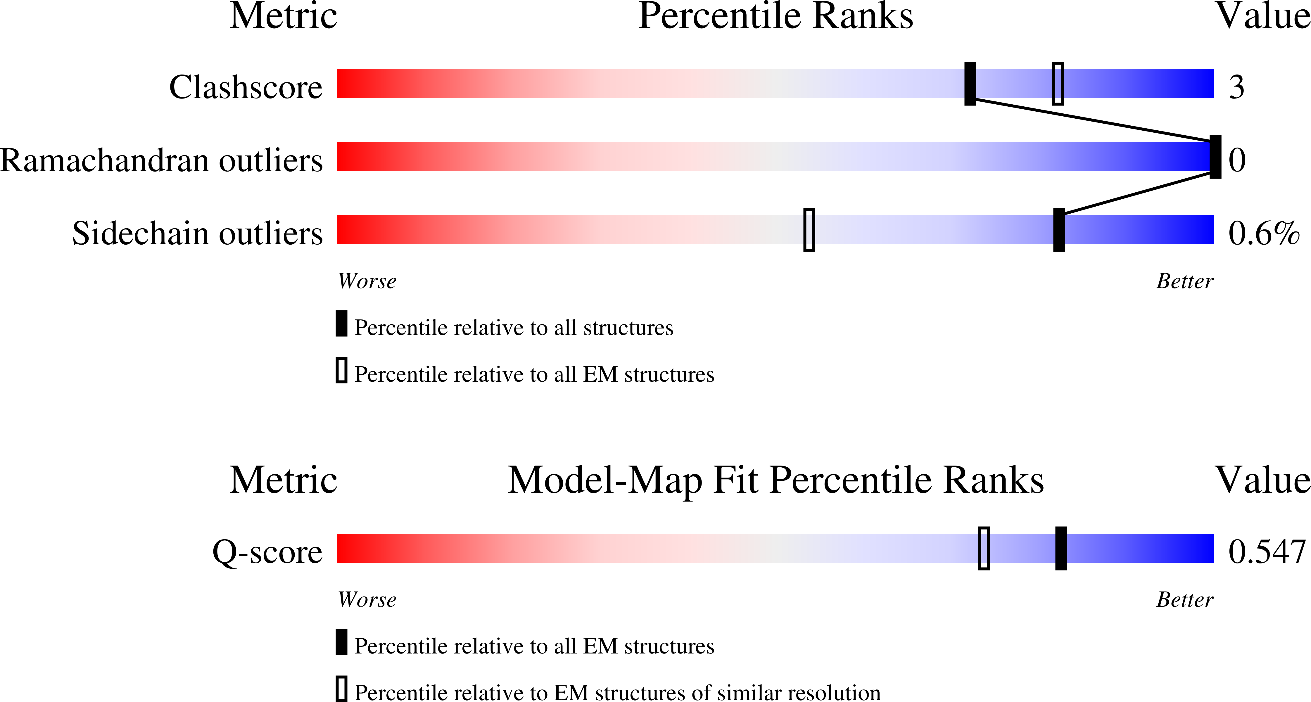
Cryo-EM structure of active human green cone opsin in complex with chimeric G protein (miniGist)
Biological Source:
Source Organism:
Homo sapiens (Taxon ID: 9606)
Mus musculus (Taxon ID: 10090)
Host Organism:
Method Details:
Experimental Method:
Resolution:
2.90 Å
Aggregation State:
PARTICLE
Reconstruction Method:
SINGLE PARTICLE
Macromolecular Entities
Polymer Type:polypeptide(L)
Molecule:Genome polyprotein,Guanine nucleotide-binding protein G(s) subunit alpha isoforms XLas
Chain IDs:E (auth: A)
Chain Length:509
Number of Molecules:1
Biological Source:Homo sapiens
Structures with similar UniProt ID
Protein Blast
Polymer Type:polypeptide(L)
Molecule:Guanine nucleotide-binding protein G(I)/G(S)/G(T) subunit beta-1
Gene (Uniprot):GNB1
Chain IDs:B
Chain Length:357
Number of Molecules:1
Biological Source:Homo sapiens
Structures with similar UniProt ID
Protein Blast
Polymer Type:polypeptide(L)
Molecule:Guanine nucleotide-binding protein G(I)/G(S)/G(O) subunit gamma-2
Gene (Uniprot):GNG2
Chain IDs:C (auth: G)
Chain Length:71
Number of Molecules:1
Biological Source:Homo sapiens
Polymer Type:polypeptide(L)
Molecule:scFv16
Chain IDs:D (auth: H)
Chain Length:259
Number of Molecules:1
Biological Source:Mus musculus
Structures with similar UniProt ID
Protein Blast
Polymer Type:polypeptide(L)
Molecule:Medium-wave-sensitive opsin 1
Gene (Uniprot):OPN1MW
Chain IDs:A (auth: R)
Chain Length:364
Number of Molecules:1
Biological Source:Homo sapiens
Ligand Molecules
Primary Citation
Structure of human green cone opsin yields insights into mechanisms underlying the rapid decay of its active, signaling state.
Proc.Natl.Acad.Sci.USA 122 e2516318122 e2516318122 (2025)
PMID: 41329744 DOI: 10.1073/pnas.2516318122

Abstact

Cone opsins enable daylight vision and color discrimination. Like their dim-light cousin rhodopsin (Rho) found in rod cells, they use a covalently attached retinal ligand to sense light and initiate visual phototransduction by activating G proteins. Unfortunately, we know less about their structural properties, in part because their activated state is unstable-cone opsins release their retinal agonist within seconds after light activation, ~100× faster than Rho. To determine what causes this rapid release and how it affects G protein activation, we solved the structure of active-state, wild-type human green cone opsin (GCOWT) stabilized with a mini-G protein and then compared its structural and biophysical properties to Rho. Our results reveal unique features in the active-state GCOWT structure. These include i) a larger water channel connected to a larger retinal binding cavity, ii) a larger "hole" near the retinal Schiff base that could facilitate both retinal escape and water access; and iii) a potential anionic residue, E102, that lies within ~3.6 Å of the Schiff base. Our biophysical assays show that neutralizing E102 (mutant GCOE102Q) slows retinal release (~8×) from the receptor and increases G protein activation. Surprisingly, our kinetic studies suggest that entropic factors are the main cause for the faster retinal release from activated GCOWT. These unique attributes in GCOWT likely facilitate its function in bright daylight. These results support the proposal that rapid retinal release from an active-state cone opsin helps prevent signal saturation and enables rapid resetting of the receptor.

Legend

Protein

Chemical

Disease

Primary Citation of related structures
Feedback Form
Name
Email
Institute
Feedback