9X2O image
Deposition Date 2025-10-07
Release Date 2025-12-03
Last Version Date 2026-01-07
Entry Detail
PDB ID:
9X2O
Title:
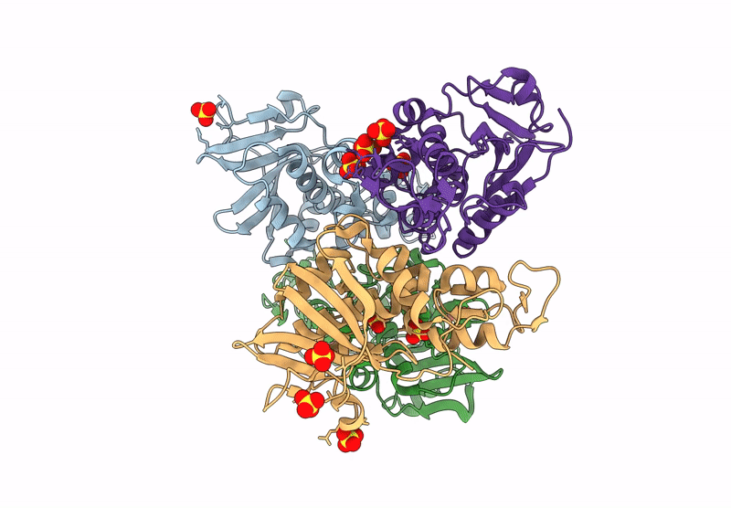
X-ray structure of antiviral protein from Mirabilis jalapa
Biological Source:
Source Organism(s):
Expression System(s):
Method Details:
Experimental Method:
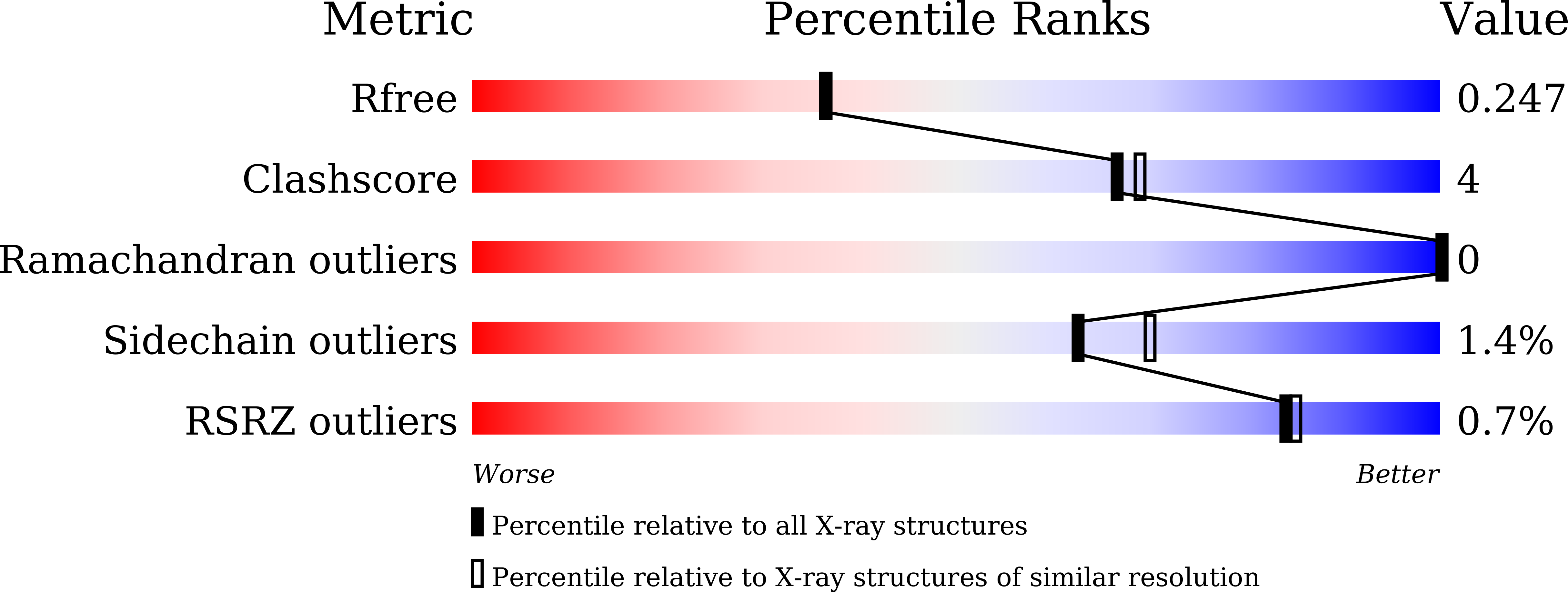
Resolution:
2.10 Å
R-Value Free:
0.24
R-Value Work:
0.19
R-Value Observed:
0.19
Space Group:
C 1 2 1
Macromolecular Entities
Structures with similar UniProt ID
Protein Blast
Polymer Type:polypeptide(L)
Molecule:Antiviral protein MAP
Chain IDs:A, B, C, D
Chain Length:256
Number of Molecules:4
Biological Source:Mirabilis jalapa
Ligand Molecules
Primary Citation
Structure of Ribosome-Inactivating Protein from Mirabilis jalapa and Its L12-Stalk-Dependent Inhibition of Escherichia coli Ribosome.
Toxins 17 ? ? (2025)
PMID: 41441611 DOI: 10.3390/toxins17120575

Abstact

Mirabilis antiviral protein (MAP) is the type I ribosome-inactivating protein (RIP), which consists of an RNA N-glycosylase domain with no carbohydrate-binding domain. Unlike many RIPs, such as ricin or trichosanthin, which inactivate eukaryotic ribosomes, MAP also inactivates the E. coli ribosome by cleaving the N-glycosidic bond at A2660 of 23S ribosomal RNA. The structure of the wild-type MAP has not been revealed yet. Here, we expressed, purified, and crystallized the plural recombinant MAPs, including both E168Q and R171Q mutations (MAP-EQRQ) in E. coli, and determined the crystal structure of MAP-EQRQ at 2.1 Å resolution. According to the predicted structure with RNA (sarcin-ricin loop) and the mutant protein's activities using quantitative RT-PCR, we showed that residue R171 at the active site of MAP is a key residue to form the stable complex with target adenine. Furthermore, we showed that MAP bound the C-terminal domains of eukaryotic P2-stalk as well as E. coli L12-stalk.

Legend

Protein

Chemical

Disease

Primary Citation of related structures
Feedback Form
Name
Email
Institute
Feedback