9VP5 image
Deposition Date 2025-07-02
Release Date 2025-11-05
Last Version Date 2025-11-05
Entry Detail
PDB ID:
9VP5
Keywords:
Title:
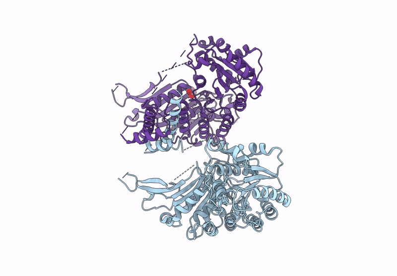
Glucose-6-phosphate dehydrogenase from Leishmania donovani
Biological Source:
Source Organism:
Method Details:
Experimental Method:
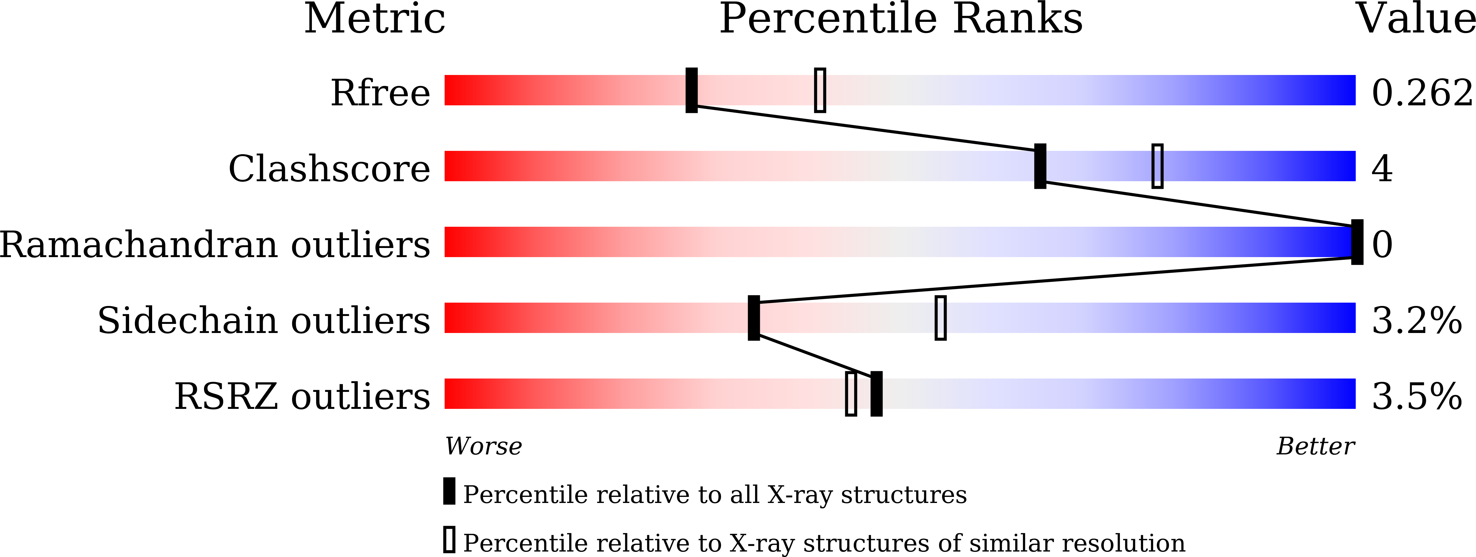
Resolution:
2.40 Å
R-Value Free:
0.25
R-Value Work:
0.19
Space Group:
C 1 2 1
Macromolecular Entities
Structures with similar UniProt ID
Protein Blast
Polymer Type:polypeptide(L)
Molecule:Glucose-6-phosphate 1-dehydrogenase
Gene (Uniprot):g6pdh
Chain IDs:A, B
Chain Length:596
Number of Molecules:2
Biological Source:Leishmania donovani
Ligand Molecules
Primary Citation
Crystal structure of leishmanial glucose-6-phosphate dehydrogenase and its implications for selective inhibitor development.
Int.J.Biol.Macromol. 331 148313 148313 (2025)
PMID: 41093198 DOI: 10.1016/j.ijbiomac.2025.148313

Abstact

Glucose-6-phosphate dehydrogenase (G6PDH) is a vital enzyme in trypanosomatid parasites, including Leishmania, where it generates NADPH to combat oxidative stress. In this study, Leishmania donovani G6PDH (LdG6PDH) was cloned, expressed, and purified to homogeneity using chromatographic methods. The purified enzyme existed as a dimer in solution, exhibited optimal activity near physiological pH, and displayed higher affinity for its cofactor (NADP) than for its substrate, glucose-6-phosphate (G6P). The enzymatic activity assay of leishmanial G6PDH revealed that ellagic acid (EA) and epigallocatechin gallate (EGCG) potently inhibit the enzyme, presenting a mixed-type mode of inhibition with respect to the substrate. Fluorescence spectroscopy established that most tryptophan residues are embedded within the hydrophobic core of LdG6PDH, while binding studies confirmed its physical interactions with ligands such as G6P, GLP, NADP, EA, and EGCG. Circular dichroism (CD) spectroscopy divulged a predominance of α-helical structure and substantiated the structural stability of the enzyme under different pH conditions. The crystal structure of LdG6PDH was resolved in both apo and ligand-bound forms, revealing three domains: a unique N-terminal domain, an NADP-binding domain, and a β + α domain. Notably, the ligand-bound structures demonstrated distinct binding positions for G6P and GLP compared to other known G6PDH structures. In silico studies identified lead compounds with a stronger affinity than NADP, indicating favorable physicochemical and pharmacokinetic properties, which were further validated through molecular dynamics simulations and MMPBSA analysis. Collectively, these findings elucidate the structural and functional properties of LdG6PDH, providing a foundation for the development of targeted inhibitors against leishmaniasis.

Legend

Protein

Chemical

Disease

Primary Citation of related structures
Feedback Form
Name
Email
Institute
Feedback