9VNJ image
Deposition Date 2025-06-30
Release Date 2025-10-08
Last Version Date 2025-10-29
Entry Detail
PDB ID:
9VNJ
Keywords:
Title:
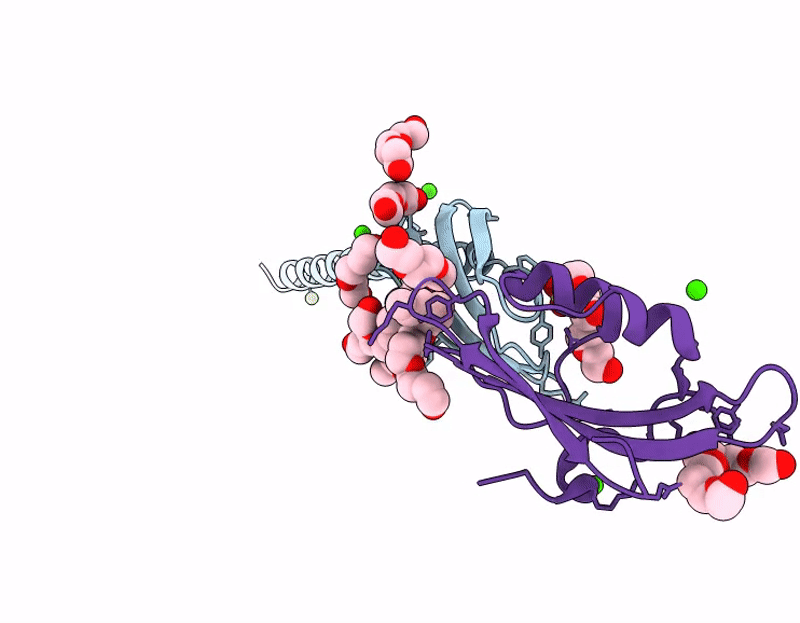
Crystal structure of the transmembrane domain of trimeric autotransporter adhesin AtaA in complex with the N-terminal domain of TpgA
Biological Source:
Source Organism:
Host Organism:
Method Details:
Experimental Method:
Resolution:
2.60 Å
R-Value Free:
0.24
R-Value Work:
0.19
R-Value Observed:
0.19
Space Group:
H 3 2
Macromolecular Entities
Structures with similar UniProt ID
Protein Blast
Polymer Type:polypeptide(L)
Molecule:Trimeric autotransporter adhesin AtaA
Gene (Uniprot):ataA
Chain IDs:A
Chain Length:108
Number of Molecules:1
Biological Source:Acinetobacter sp. Tol 5
Structures with similar UniProt ID
Protein Blast
Polymer Type:polypeptide(L)
Molecule:Trimeric autotransporter adhesin- and peptidoglycan-associated protein A
Gene (Uniprot):tpgA
Chain IDs:B
Chain Length:127
Number of Molecules:1
Biological Source:Acinetobacter sp. Tol 5
Primary Citation
Insights into the complex formation of a trimeric autotransporter adhesin with a peptidoglycan-binding periplasmic protein.
Cell Surf 14 100155 100155 (2025)
PMID: 41103730 DOI: 10.1016/j.tcsw.2025.100155

Abstact

Trimeric autotransporter adhesins (TAAs) are outer membrane (OM) proteins that are widely distributed in gram-negative bacteria and are involved primarily in adhesion to biotic and abiotic surfaces, cell agglutination, and biofilm formation. TAAs consist of a passenger domain, which is secreted onto the cell surface, and a transmembrane domain, which forms a pore in the OM to secrete and anchor the passenger domain. Because the interactions between TAAs and chaperones or dedicated auxiliary proteins during secretion are short-lived, TAAs are thought to reside on the OM without forming complexes with other proteins after secretion. In this study, we aimed to clarify the interactions between an Acinetobacter TAA, AtaA, and a peptidoglycan (PG)-binding periplasmic protein, TpgA. Pull-down assays using recombinant proteins identified the interacting domains. X-ray crystallography at 2.6 Å resolution revealed an A3B3 heterohexameric complex structure composed of the N-terminal domain of TpgA and the transmembrane domain of AtaA. TpgA-N consists of two short α helices and three antiparallel β strands, yielding an ααβββ topology similar to BamE. However, the regions corresponding to BamE interfaces with BamA and BamD differ in TpgA-N. All-atom molecular dynamics simulations and mutational assays revealed that both electrostatic and hydrophobic interactions contribute to stable complex formation. Bioinformatic analyses indicate that the TAA-TpgA complex occurs in a wide range of species. These findings will contribute to a better understanding of TAAs and the cell envelope.

Legend

Protein

Chemical

Disease

Primary Citation of related structures