9VGW image
Deposition Date 2025-06-15
Release Date 2025-09-10
Last Version Date 2025-11-19
Entry Detail
PDB ID:
9VGW
Title:
Crystal structure of human PCNA in complex with REV1 PIP-box
Biological Source:
Source Organism:
Homo sapiens (Taxon ID: 9606)
Host Organism:
Method Details:
Experimental Method:
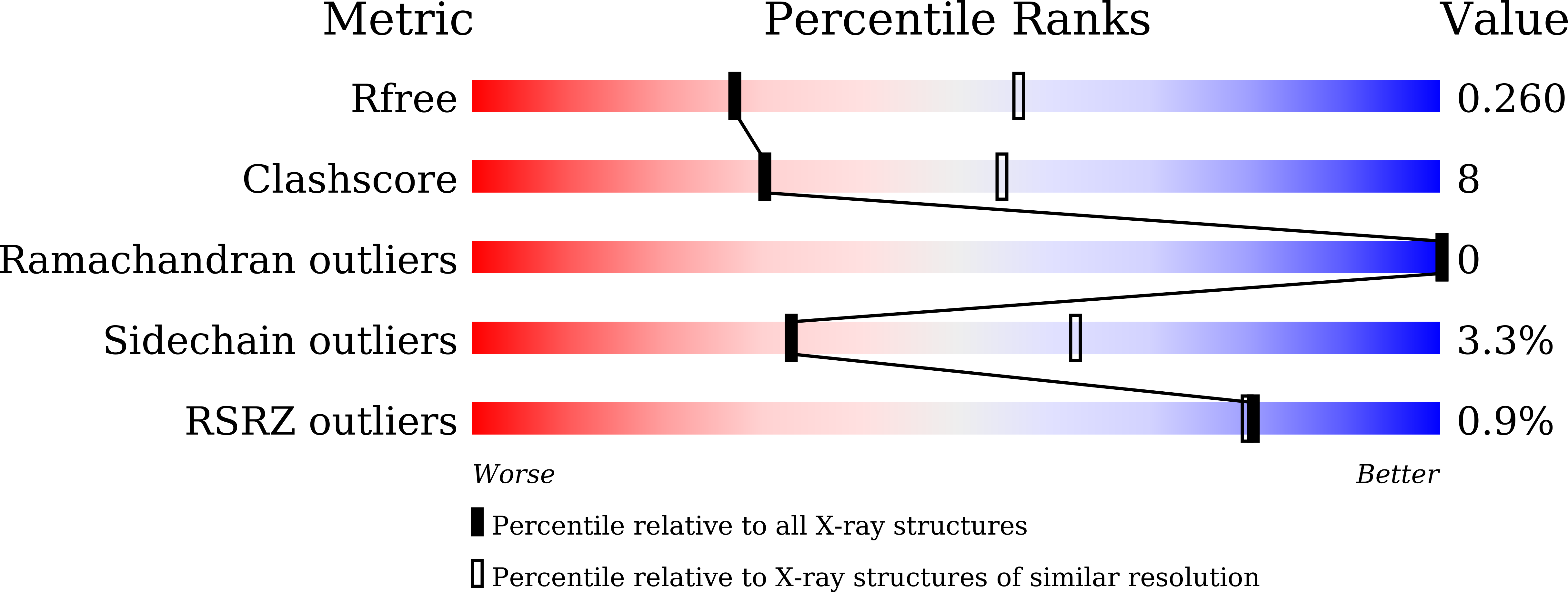
Resolution:
2.70 Å
R-Value Free:
0.26
R-Value Work:
0.21
R-Value Observed:
0.21
Space Group:
P 21 21 2
Macromolecular Entities
Structures with similar UniProt ID
Protein Blast
Polymer Type:polypeptide(L)
Molecule:Proliferating cell nuclear antigen
Gene (Uniprot):PCNA
Chain IDs:A, B, C
Chain Length:261
Number of Molecules:3
Biological Source:Homo sapiens
Structures with similar UniProt ID
Protein Blast
Polymer Type:polypeptide(L)
Molecule:peptide from DNA repair protein REV1
Gene (Uniprot):REV1
Chain IDs:D (auth: X), E (auth: Y), F (auth: Z)
Chain Length:13
Number of Molecules:3
Biological Source:Homo sapiens
Primary Citation
Identification of a PCNA-binding motif in human translesion DNA polymerase REV1 and structural basis of its interaction with PCNA.
J.Biochem. 178 315 324 (2025)
PMID: 40888629 DOI: 10.1093/jb/mvaf054

Abstact

REV1 is a eukaryotic error-prone DNA polymerase belonging to the Y-family, with a central role in translesion DNA synthesis (TLS) to continue DNA replication even in the presence of DNA damage in the template strand. TLS is stimulated by monoubiquitination of proliferating cell nuclear antigen (PCNA), a toroidal-shaped protein functioning as a scaffold for DNA polymerases and repair enzymes. Mammals possess four types of Y-family DNA polymerases: Pol η, Pol κ, Pol ι and REV1. Among those, Pol η, Pol κ and Pol ι interact with PCNA through PCNA-binding motifs, low-affinity variants of PCNA-interacting protein box (PIP-box). To date, several studies have reported that REV1 interacts with PCNA, but identified PCNA-binding regions are inconsistent; therefore, a structural basis for interaction between REV1 and PCNA also remains unclear. Here, we identified a signature sequence conserved within vertebrates REV1 responsible for PCNA-binding. Furthermore, we unveiled a mechanism underlying the physical interaction between the PCNA-binding motif of human REV1 and PCNA by X-ray crystallography, thus revealing that REV1 binds to PCNA through a PIP-box variant located in the C-terminal side of the little finger domain. Our study provides a convincing answer for a long-standing controversy regarding the physical interaction between REV1 and PCNA.

Legend

Protein

Chemical

Disease

Primary Citation of related structures
Feedback Form
Name
Email
Institute
Feedback