9VF8 image
Deposition Date 2025-06-10
Release Date 2025-06-25
Last Version Date 2025-12-17
Entry Detail
PDB ID:
9VF8
Title:
Structure of Meiothermus ruber Mrub_1259 LOV domain (MrLOV)
Biological Source:
Source Organism(s):
Expression System(s):
Method Details:
Experimental Method:
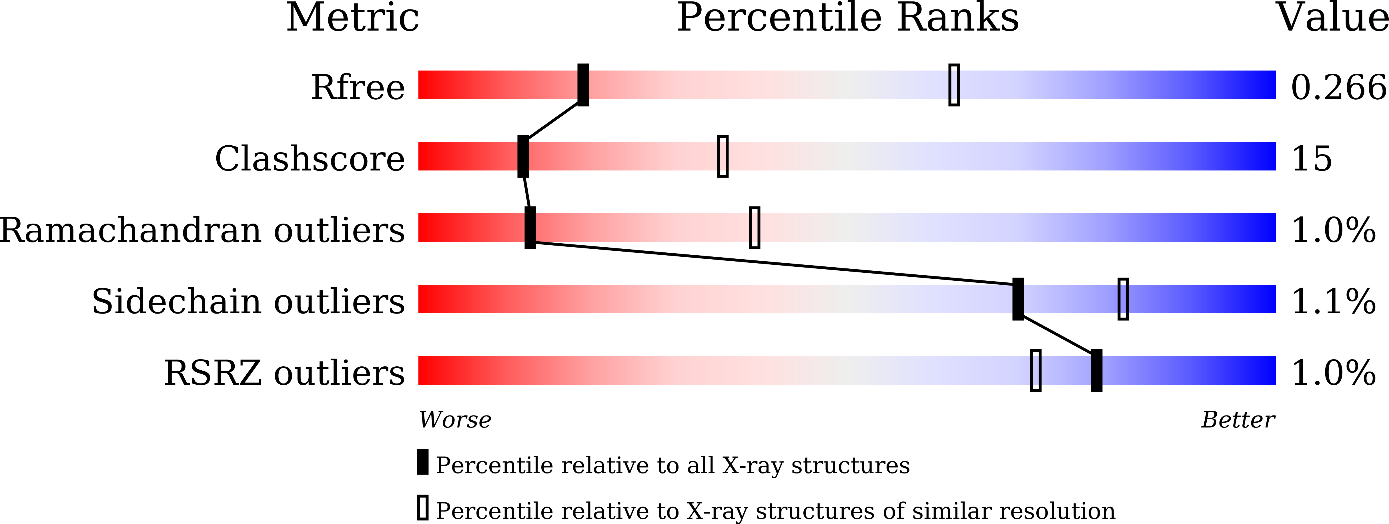
Resolution:
3.35 Å
R-Value Free:
0.27
R-Value Work:
0.24
Space Group:
P 43 21 2
Macromolecular Entities
Structures with similar UniProt ID
Protein Blast
Polymer Type:polypeptide(L)
Molecule:histidine kinase
Gene (Uniprot):K649_05955
Chain IDs:A
Chain Length:112
Number of Molecules:1
Biological Source:Meiothermus ruber DSM 1279
Ligand Molecules
Primary Citation
Structural characterization of Meiothermus ruber LOV domain.
J.Struct.Biol. 218 108268 108268 (2025)
PMID: 41349808 DOI: 10.1016/j.jsb.2025.108268

Abstact

Light Oxygen Voltage (LOV) domains are important widespread receptors of blue light that also found applications in optogenetics and imaging. While LOV domains from mesophiles are relatively well characterized, their counterparts from thermophilic microorganisms remain understudied. Here, we express two constructs of a LOV domain belonging to a histidine kinase from Meiothermus ruber, MrLOV and MrLOVe, and show that they are photoactive, with recovery time values of 21 and 27 min, respectively, and thermostable. Crystal structures reveal that MrLOV, which lacks helices A'α and Jα, forms a parallel dimer, whereas MrLOVe is a tetramer organized as an antiparallel dimer of two parallel dimers interacting via helices Jα. One MrLOVe dimer is symmetric, and the other is asymmetric, with conformational differences mirroring activation-related changes in other LOV domains. Our data provide the structural basis for understanding and engineering of thermophilic LOVs and pave the way for development of thermostable and photostable LOV-derived optogenetic tools and flavin-based fluorescent proteins.

Legend

Protein

Chemical

Disease

Primary Citation of related structures
Feedback Form
Name
Email
Institute
Feedback