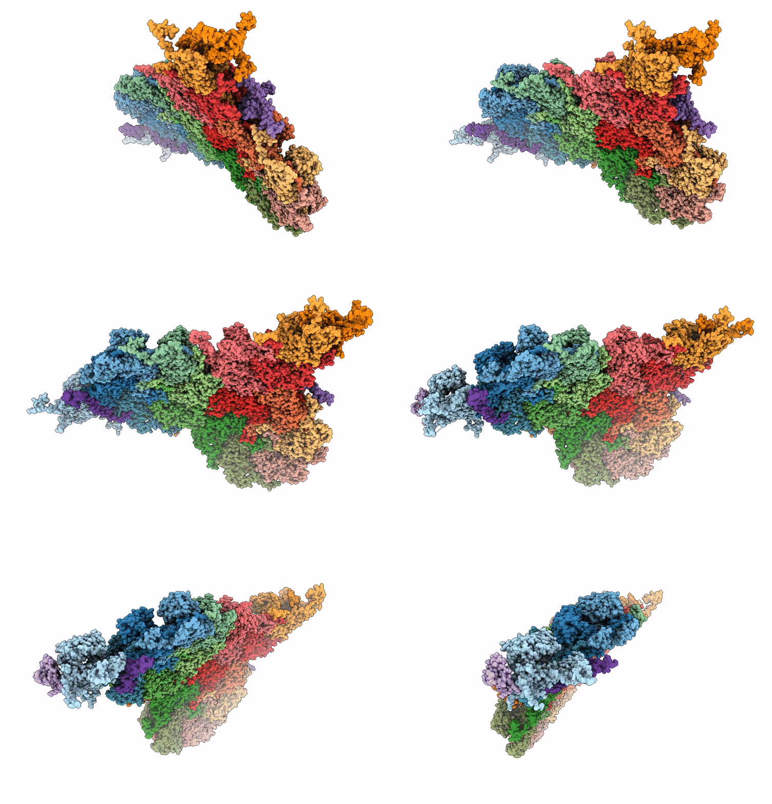
9VEL image
Deposition Date 2025-06-09
Release Date 2025-08-27
Last Version Date 2025-09-10
Entry Detail
PDB ID:
9VEL
Keywords:
Title:
The composite cryo-EM structure of bacteriophage SPO1 capsid
Biological Source:
Source Organism:
Method Details:
Experimental Method:
Resolution:
3.00 Å
Aggregation State:
PARTICLE
Reconstruction Method:
SINGLE PARTICLE
Macromolecular Entities
Structures with similar UniProt ID
Protein Blast
Polymer Type:polypeptide(L)
Molecule:Gp6.1
Gene (Uniprot):6.1
Chain IDs:A (auth: C), B (auth: F), C (auth: G), D, E (auth: I), F (auth: J), G (auth: O), H (auth: N), I (auth: M), J (auth: H), K, L, M (auth: B), Q (auth: E), R (auth: P), S (auth: A)
Chain Length:468
Number of Molecules:16
Biological Source:Bacillus phage SPO1
Structures with similar UniProt ID
Protein Blast
Polymer Type:polypeptide(L)
Molecule:Gp29.2
Gene (Uniprot):29.2
Chain IDs:N (auth: r), O (auth: q), P (auth: s), U (auth: v)
Chain Length:171
Number of Molecules:4
Biological Source:Bacillus phage SPO1
Structures with similar UniProt ID
Protein Blast
Polymer Type:polypeptide(L)
Molecule:Gp2.7
Gene (Uniprot):2.7
Chain IDs:T (auth: t)
Chain Length:57
Number of Molecules:1
Biological Source:Bacillus phage SPO1
Structures with similar UniProt ID
Protein Blast
Polymer Type:polypeptide(L)
Molecule:Gp36.3
Gene (Uniprot):36.3
Chain IDs:V (auth: w)
Chain Length:96
Number of Molecules:1
Biological Source:Bacillus phage SPO1
Ligand Molecules
Primary Citation
Capsid structure of phage SPO1 reveals novel minor capsid proteins and insights into capsid stabilization.
Structure ? ? ? (2025)
PMID: 40885197 DOI: 10.1016/j.str.2025.08.004

Abstact

SPO1-related bacteriophages are promising candidates for phage therapy. We present the 3.0 Å cryo-electron microscopy (cryo-EM) structure of the SPO1 capsid with a triangulation number T = 16, enabling the construction of an atomic model comprising the major capsid protein and three types of minor capsid proteins: gp29.2, gp2.7, and gp36.3. These minor capsid proteins adopt novel folds. They might stabilize the capsid and determine its curvature. Gp29.2 monomers contain a three-blade propeller fold and are located at the 3-fold and quasi-three-fold axes. Gp2.7 forms pentamers atop pentameric capsomers, while gp36.3 binds to the capsid's inner surface, forming star-shaped structures increasing connections between pentameric and hexameric capsomers. The surface exposed regions of gp29.2 and gp2.7 make SPO1 of interest as a nanocage for phage display. Our findings advance the understanding of capsid architecture, stabilization, and local curvature determination for SPO1-related bacteriophages.

Legend

Protein

Chemical

Disease

Primary Citation of related structures
Feedback Form
Name
Email
Institute
Feedback