9VBK image
Deposition Date 2025-06-04
Release Date 2025-12-24
Last Version Date 2025-12-24
Entry Detail
PDB ID:
9VBK
Keywords:
Title:
Cryo-EM structure of human PLD4 bound to ssDNA (poly(T))
Biological Source:
Source Organism:
Host Organism:
Method Details:
Experimental Method:
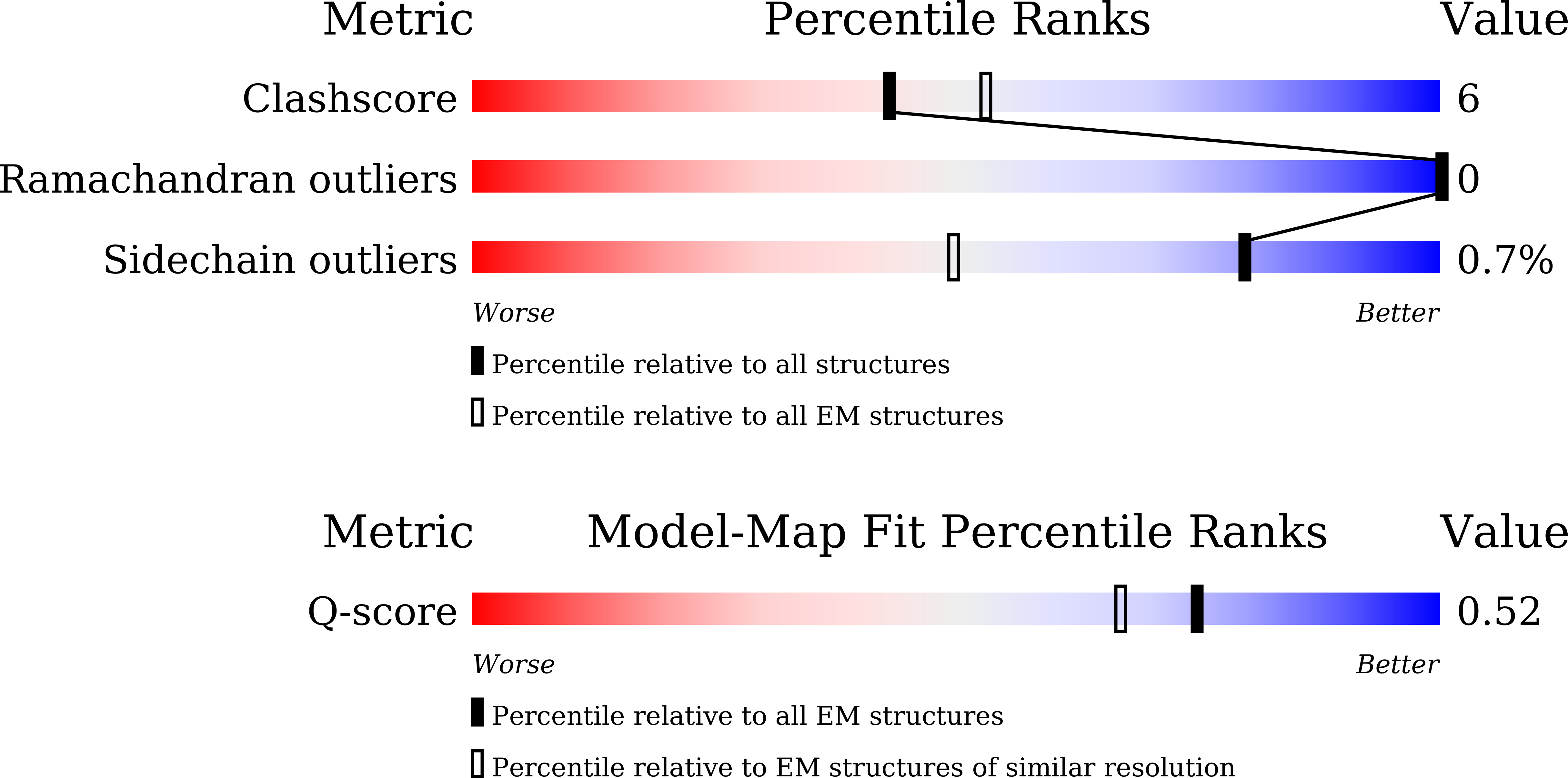
Resolution:
3.06 Å
Aggregation State:
PARTICLE
Reconstruction Method:
SINGLE PARTICLE
Macromolecular Entities
Structures with similar UniProt ID
Protein Blast
Polymer Type:polypeptide(L)
Molecule:5'-3' exonuclease PLD4
Gene (Uniprot):PLD4
Chain IDs:A, B, C, D
Chain Length:422
Number of Molecules:4
Biological Source:Homo sapiens
Polymer Type:polydeoxyribonucleotide
Molecule:DNA (5'-D(*TP*TP*T)-3')
Chain IDs:E (auth: F), F (auth: G)
Chain Length:49
Number of Molecules:2
Biological Source:synthetic construct
Primary Citation
Mechanistic insights into single-stranded DNA degradation by lysosomal exonucleases PLD3 and PLD4 from structural snapshots.
Nat Commun ? ? ? (2025)
PMID: 41381514 DOI: 10.1038/s41467-025-66261-2

Abstact

Lysosomal exonuclease phospholipase D (PLD) family PLD3 and PLD4 degrade single-stranded RNA or DNA and regulate TLR7 or TLR9 responses. Polymorphisms of these enzymes are associated with human diseases: PLD4 is associated with inflammatory diseases, and PLD3 is associated with neurodegenerative diseases. Here, we determine the structures of substrate-bound PLD3 and PLD4 by cryo-electron microscopy. Our structures reveal that PLD3 rebuilds a substrate-binding pocket, depending on the substrate, mainly via motion of the Phe335-containing loop. Furthermore, we captured the structure in a metastable state that appears during substrate rearrangement following product release. Together, our findings identify the residues that underlie the distinct activities of PLD3 and PLD4. This study provides a mechanistic basis for the exonuclease activity of PLD3 and PLD4 in single-stranded DNA degradation.

Legend

Protein

Chemical

Disease

Primary Citation of related structures
Feedback Form
Name
Email
Institute
Feedback