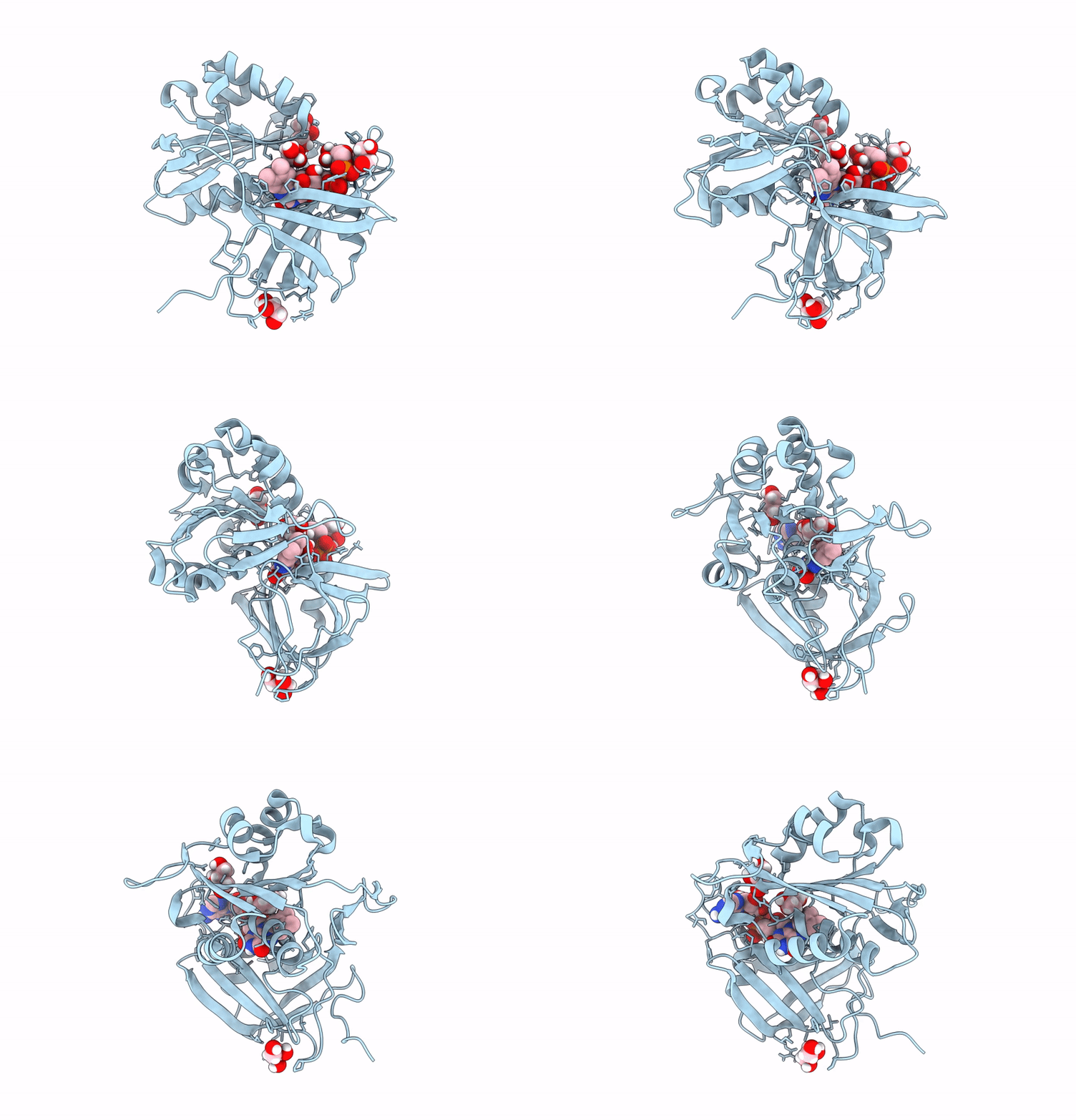
9V6C image
Deposition Date 2025-05-27
Release Date 2025-11-19
Last Version Date 2025-11-19
Entry Detail
PDB ID:
9V6C
Keywords:
Title:
Neutron crystal structure of the oxidized form of b5R at pD 7.5
Biological Source:
Source Organism:
Sus scrofa (Taxon ID: 9823)
Host Organism:
Method Details:
Experimental Method:
Resolution:
1.45 Å
R-Value Free:
0.20
R-Value Work:
0.17
R-Value Observed:
0.17
Space Group:
P 21 21 21
Macromolecular Entities
Structures with similar UniProt ID
Protein Blast
Polymer Type:polypeptide(L)
Molecule:NADH-cytochrome b5 reductase 3
Gene (Uniprot):CYB5R3
Chain IDs:A
Chain Length:272
Number of Molecules:1
Biological Source:Sus scrofa
Primary Citation
Structural basis of hydride and proton transfer reactions revealed by the detection of hydrogen atoms in mammalian NADH-cytochrome b 5 reductase.
Structure ? ? ? (2025)
PMID: 41172987 DOI: 10.1016/j.str.2025.10.006

Abstact

Many structural studies have been reported for ferredoxin:NADP+ reductase family members, but an experimental validation of the catalytic hydride and proton transfer steps through a direct detection of the involved hydrogen atoms has not been achieved so far. Here, we determined high-resolution X-ray and neutron crystal structures of NADH-cytochrome b5 reductase, which acts as an electron supplier for various metabolic processes and mediates hydride and proton transfer reactions via its FAD and NADH cofactors. The X-ray structures identify the FADH--NAD+ and FAD-NADH complexes based on the electron densities of the hydrogen atoms bound to the cofactors. The neutron structures determined at different pD-values show a difference in the protonation state of the histidine residue in the hydrogen-bond network from FAD to the protein surface. The observation of the hydrogen atoms reveals the structural basis for the hydride and proton transfer reactions catalyzed by NADH-cytochrome b5 reductase.

Legend

Protein

Chemical

Disease

Primary Citation of related structures
Feedback Form
Name
Email
Institute
Feedback