9UWZ image
Deposition Date 2025-05-13
Release Date 2025-06-25
Last Version Date 2025-07-02
Entry Detail
PDB ID:
9UWZ
Keywords:
Title:
Crystal structure of the type III secretion chaperone VecA from Vibrio parahaemolyticus
Biological Source:
Source Organism:
Host Organism:
Method Details:
Experimental Method:
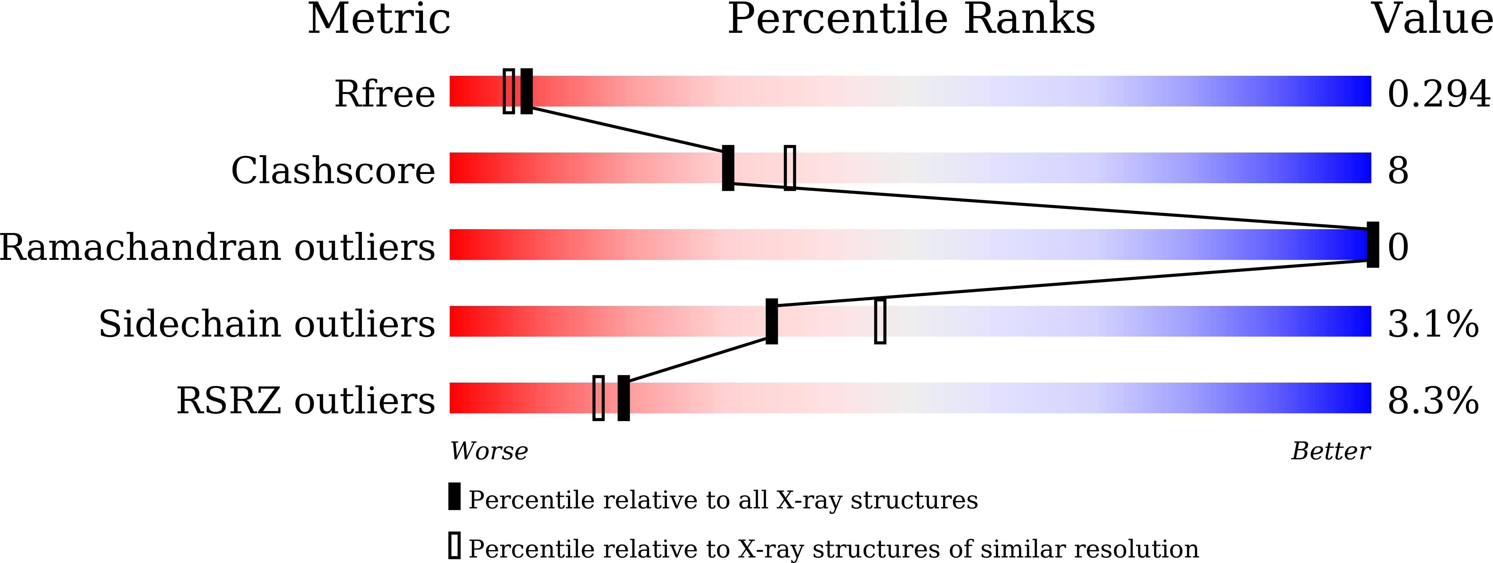
Resolution:
2.20 Å
R-Value Free:
0.29
R-Value Work:
0.23
R-Value Observed:
0.24
Space Group:
P 41 21 2
Macromolecular Entities
Polymer Type:polypeptide(L)
Molecule:CesT family type III secretion system chaperone
Gene (Uniprot):VP1682
Chain IDs:A, B
Chain Length:157
Number of Molecules:2
Biological Source:Vibrio parahaemolyticus
Primary Citation
Structural basis of effector recognition by the T3SS chaperone VecA from Vibrio parahaemolyticus.
Biochem.Biophys.Res.Commun. 776 152190 152190 (2025)
PMID: 40517674 DOI: 10.1016/j.bbrc.2025.152190

Abstact

Many pathogenic gram-negative bacteria utilise the type III secretion system (T3SS), a specific protein injection apparatus, to translocate virulence effectors into host cells, modulating host cell functions and establishing infection. To facilitate the precise cytosolic transport of effectors to T3SS, a class of proteins called chaperones plays a crucial role. However, a limited number of available structural data on chaperone-effector complexes hampers understanding of the mechanisms underlying this process. In Vibrio parahaemolyticus, a major causative agent of seafood-associated acute gastroenteritis in humans, T3SS chaperone VecA transports its cognate membrane-disrupting effector, VepA. Here, we determined the crystal structure of VecA alone and in complex with VepA at resolutions of 2.20 Å and 2.49 Å, respectively. While the overall protein fold and the hydrophobic cleft that accommodates an N-terminal β-motif of effectors were conserved among T3SS chaperones, the structural analysis revealed that surface residues are remarkably different, reflecting their substrate specificity. Additionally, unlike other reported structures of the T3SS chaperone-effector complexes, in which the effectors are partially unfolded and wrapped around the chaperone, VepA adopts a highly folded conformation in the complex. This compact structure appears to protect the fragile glycine-rich transmembrane domain of VepA and suggests that upon secretion, VepA undergoes conformational changes, including α-helix formation, allowing the transmembrane domain to embed into and disrupt the membrane of organelles containing its binding target, V-ATPase. These findings elucidate the chaperone-mediated regulation of effector transport and function of the bacterial virulence-related T3SS.

Legend

Protein

Chemical

Disease

Primary Citation of related structures