9UAJ image
Deposition Date 2025-04-01
Release Date 2025-12-31
Last Version Date 2025-12-31
Entry Detail
PDB ID:
9UAJ
Title:
Ovorubin from the golden apple snail (Pomacea canaliculata)
Biological Source:
Source Organism:
Method Details:
Experimental Method:
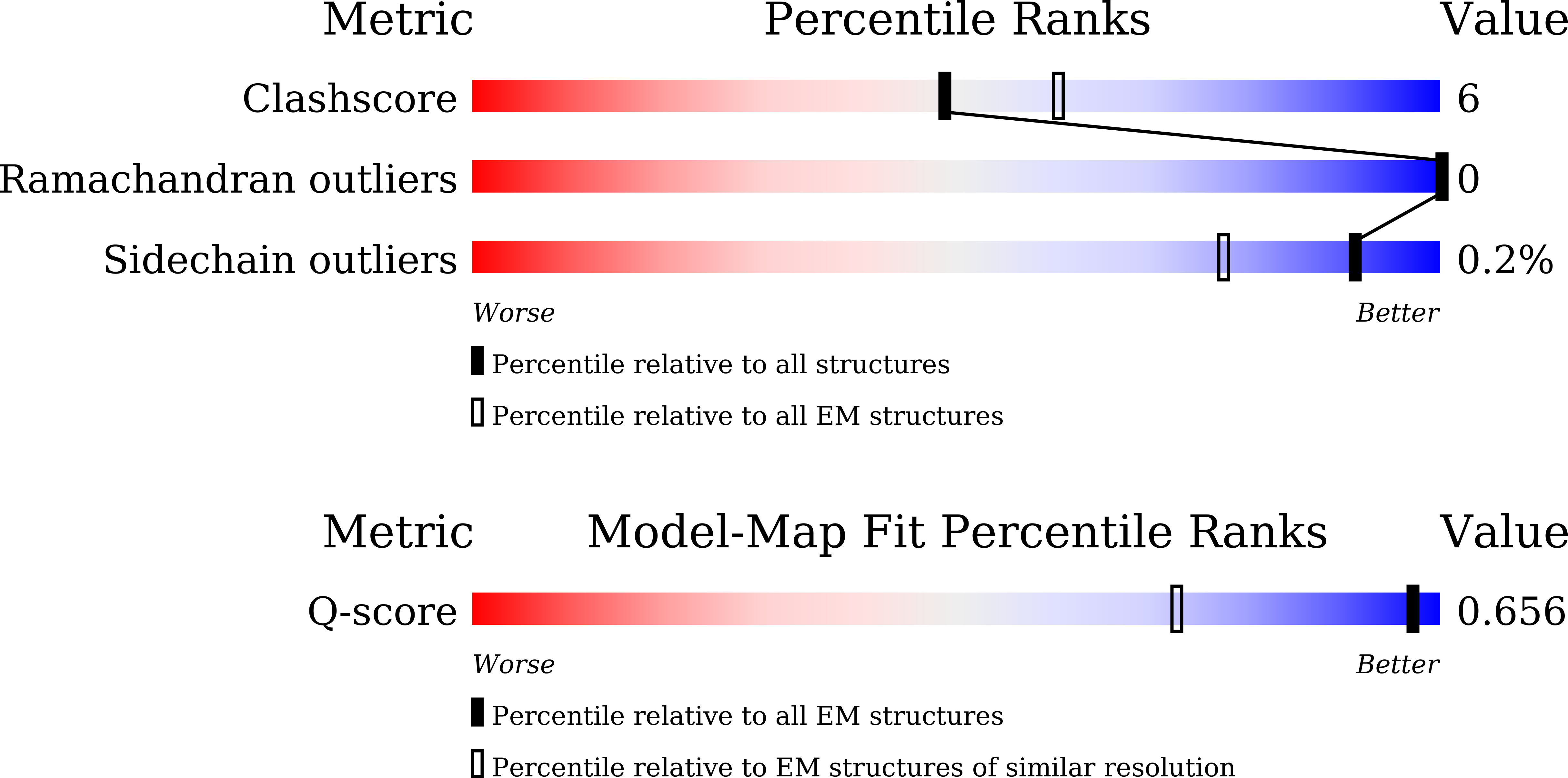
Resolution:
2.12 Å
Aggregation State:
PARTICLE
Reconstruction Method:
SINGLE PARTICLE
Macromolecular Entities
Structures with similar UniProt ID
Protein Blast
Polymer Type:polypeptide(L)
Molecule:PcOvo5
Gene (Uniprot):C0Q70_15738
Chain IDs:A, F
Chain Length:204
Number of Molecules:2
Biological Source:Pomacea canaliculata
Structures with similar UniProt ID
Protein Blast
Polymer Type:polypeptide(L)
Molecule:PcOvo3s
Gene (Uniprot):C0Q70_15738
Chain IDs:B, J
Chain Length:199
Number of Molecules:2
Biological Source:Pomacea canaliculata
Structures with similar UniProt ID
Protein Blast
Polymer Type:polypeptide(L)
Molecule:Perivitellin ovorubin-1
Gene (Uniprot):Ovo1
Chain IDs:C, I
Chain Length:200
Number of Molecules:2
Biological Source:Pomacea canaliculata
Structures with similar UniProt ID
Protein Blast
Polymer Type:polypeptide(L)
Molecule:Perivitellin protein
Gene (Uniprot):C0Q70_15737
Chain IDs:D, H
Chain Length:203
Number of Molecules:2
Biological Source:Pomacea canaliculata
Structures with similar UniProt ID
Protein Blast
Polymer Type:polypeptide(L)
Molecule:Perivitellin ovorubin-2
Gene (Uniprot):Ovo2
Chain IDs:E, G
Chain Length:210
Number of Molecules:2
Biological Source:Pomacea canaliculata
Primary Citation
Structure of the chromoprotein ovorubin from the golden apple snail (Pomacea canaliculata).
Protein Sci. 35 e70439 e70439 (2026)
PMID: 41432377 DOI: 10.1002/pro.70439

Abstact

The golden apple snail (Pomacea canaliculata), an invasive gastropod, produces distinctly bright pink egg masses. The astaxanthin-binding ovorubin (P. canaliculata ovorubin [PcOvo]) is a 300-kDa glycoprotein responsible for the egg coloration. Here, we determine the three-dimensional structure of PcOvo using cryo-electron microscopy (cryo-EM). PcOvo is a heterodecameric protein consisting of two copies of each PcOvo1-5 subunit. The subunits have similar repeated ferredoxin-like structures. N-linked glycosylation sites are identified. Solution x-ray scattering data support the overall architecture of the complex. PcOvo3 and PcOvo5 have hydrophobic pockets that likely bind various hydrophobic compounds. The cryo-EM map and binding experiments suggest that PcOvo5 binds astaxanthin, which is responsible for the pink color of the protein. Our structure provides molecular insights into the nature of the gastropod egg coloration and lays a foundation for further investigation of ovorubin biology and evolution.

Legend

Protein

Chemical

Disease

Primary Citation of related structures
Feedback Form
Name
Email
Institute
Feedback