9U81 image
Deposition Date 2025-03-25
Release Date 2025-12-10
Last Version Date 2025-12-10
Entry Detail
PDB ID:
9U81
Title:
Cryo-EM structure of tolvaptan-bound human vasopressin V2 receptor complex with Fab
Biological Source:
Source Organism:
Homo sapiens (Taxon ID: 9606)
Escherichia coli (Taxon ID: 562)
Host Organism:
Method Details:
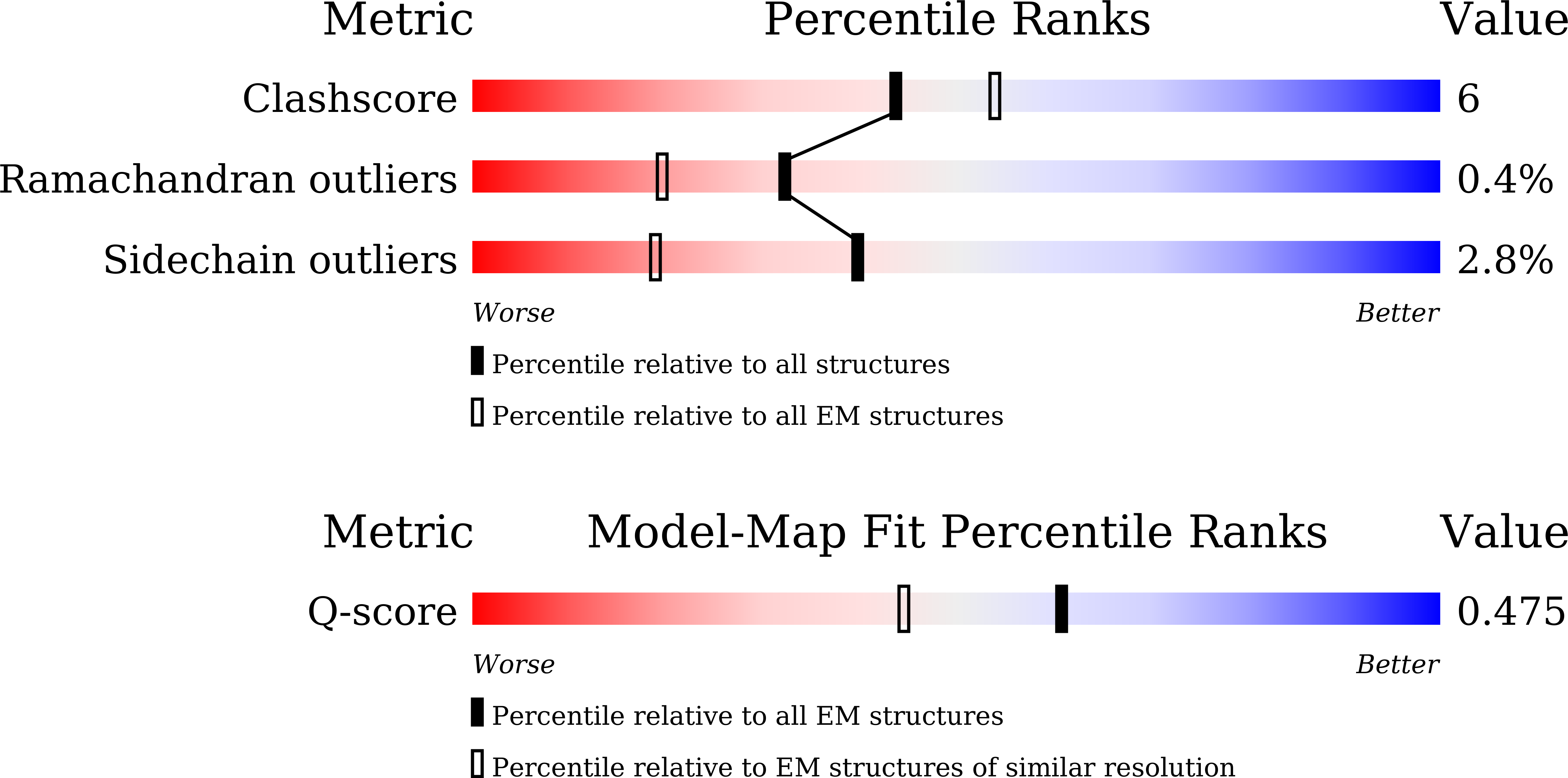
Experimental Method:
Resolution:
3.08 Å
Aggregation State:
PARTICLE
Reconstruction Method:
SINGLE PARTICLE
Macromolecular Entities
Polymer Type:polypeptide(L)
Molecule:anti-BRIL Fab heavy chain
Chain IDs:A (auth: H)
Chain Length:229
Number of Molecules:1
Biological Source:Homo sapiens
Polymer Type:polypeptide(L)
Molecule:anti-BRIL Fab light chain
Chain IDs:B (auth: L)
Chain Length:216
Number of Molecules:1
Biological Source:Homo sapiens
Protein Blast
Polymer Type:polypeptide(L)
Molecule:Vasopressin V2 receptor,Soluble cytochrome b562
Gene (Uniprot):cybC, AVPR2
Chain IDs:C (auth: R)
Chain Length:460
Number of Molecules:1
Biological Source:Homo sapiens, Escherichia coli
Ligand Molecules
Primary Citation
Structural insights into antagonist recognition by the vasopressin V2 receptor.
Nat Commun 16 9734 9734 (2025)
PMID: 41188237 DOI: 10.1038/s41467-025-64735-x

Abstact

The vasopressin V2 receptor (V2R), a class A G protein-coupled receptor, is essential for regulating body water homeostasis. V2R antagonists have emerged as promising treatments for hyponatremia; however, the absence of structural information for antagonist-bound V2R hampers our understanding of antagonist recognition and the targeted design of V2R antagonists. In this study, we present two cryo-electron microscopy structures of inactive V2R bound to the clinically approved antagonists tolvaptan and conivaptan. Combined with functional analyses and molecular dynamic simulations, these structures reveal distinct binding poses: tolvaptan is deeply inserted within the binding pocket, whereas conivaptan is positioned at a shallower depth. Integrated analyses further define critical pharmacophoric features governing antagonist activity and unveil a TM7 helical conformation-dependent antagonism mechanism that is distinct from classical GPCR inactivation modes. Our findings deepen understanding of antagonist recognition and antagonism of V2R, providing a foundation for the development of V2R-targeted therapies.

Legend

Protein

Chemical

Disease

Primary Citation of related structures
Feedback Form
Name
Email
Institute
Feedback