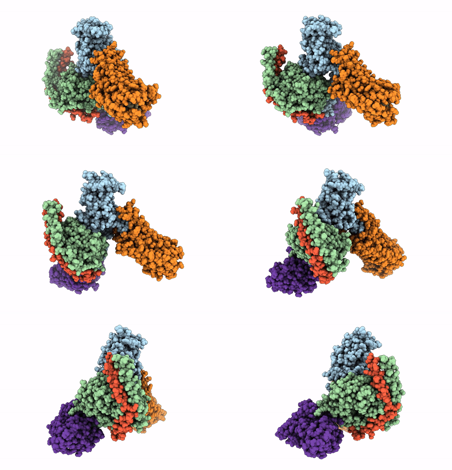
9U4W image
Deposition Date 2025-03-20
Release Date 2025-06-11
Last Version Date 2025-07-02
Entry Detail
PDB ID:
9U4W
Title:
cryo-EM structure of pig GnRHR bound with mammal GnRH
Biological Source:
Source Organism:
Homo sapiens (Taxon ID: 9606)
Sus scrofa (Taxon ID: 9823)
Host Organism:
Method Details:
Experimental Method:
Resolution:
3.18 Å
Aggregation State:
PARTICLE
Reconstruction Method:
SINGLE PARTICLE
Macromolecular Entities
Polymer Type:polypeptide(L)
Molecule:Guanine nucleotide-binding protein G(q) subunit alpha-1
Chain IDs:A
Chain Length:246
Number of Molecules:1
Biological Source:Homo sapiens
Polymer Type:polypeptide(L)
Molecule:Guanine nucleotide-binding protein G(I)/G(S)/G(T) subunit beta-1
Gene (Uniprot):GNB1
Chain IDs:B
Chain Length:344
Number of Molecules:1
Biological Source:Homo sapiens
Polymer Type:polypeptide(L)
Molecule:Gonadoliberin-1
Gene (Uniprot):GNRH1
Chain IDs:C (auth: F)
Chain Length:11
Number of Molecules:1
Biological Source:Homo sapiens
Polymer Type:polypeptide(L)
Molecule:Guanine nucleotide-binding protein G(I)/G(S)/G(O) subunit gamma-2
Gene (Uniprot):GNG2
Chain IDs:D (auth: G)
Chain Length:71
Number of Molecules:1
Biological Source:Homo sapiens
Polymer Type:polypeptide(L)
Molecule:Beta-2 adrenergic receptor,Gonadotropin-releasing hormone receptor
Gene (Uniprot):GNRHR
Chain IDs:E (auth: R)
Chain Length:414
Number of Molecules:1
Biological Source:Homo sapiens, Sus scrofa
Polymer Type:polypeptide(L)
Molecule:scFv16
Chain IDs:F (auth: S)
Chain Length:285
Number of Molecules:1
Biological Source:Homo sapiens
Modified Residue
Compound ID Chain ID Parent Comp ID Details 2D Image
PCA C GLN modified residue
Ligand Molecules
Primary Citation
Cryo-EM structures of GnRHR: Foundations for next-generation therapeutics.
Proc.Natl.Acad.Sci.USA 122 e2500112122 e2500112122 (2025)
PMID: 40523184 DOI: 10.1073/pnas.2500112122

Abstact

Gonadotropin-releasing hormone receptor (GnRHR) is critical for reproductive health and a key therapeutic target for endocrine disorders and hormone-responsive cancers. Using high-resolution cryoelectron microscopy, we determined the structures of Sus scrofa and Xenopus laevis GnRHRs bound to mammal GnRH, uncovering conserved and species-specific mechanisms of receptor activation and G protein coupling. The conserved "U"-shaped GnRH conformation mediates high-affinity binding through key interactions with residues such as K3.32, Y6.51, and Y6.52. Species-specific variations in extracellular loops and receptor-ligand contacts fine-tune receptor function, while ligand binding induces structural rearrangements, including N terminus displacement and TM6 rotation, critical for signaling. Structure-activity relationship analysis demonstrates how D-amino acid substitutions in GnRH analogs enhance stability and receptor affinity. Distinct binding modes of agonists and antagonists elucidate mechanisms of ligand-dependent activation and inactivation. These insights lay the groundwork for designing next-generation GnRHR therapeutics with enhanced specificity and efficacy for conditions like endometriosis, prostate cancer, and infertility.

Legend

Protein

Chemical

Disease

Primary Citation of related structures