9SDX image
Deposition Date 2025-08-15
Release Date 2025-12-24
Last Version Date 2026-01-14
Entry Detail
PDB ID:
9SDX
Keywords:
Title:
Structure of RBR binding E2 variant crosslinked with NEDD8-CUL5-RBX2 bound ARIH2 and Ub
Biological Source:
Source Organism(s):
Expression System(s):
Method Details:
Experimental Method:
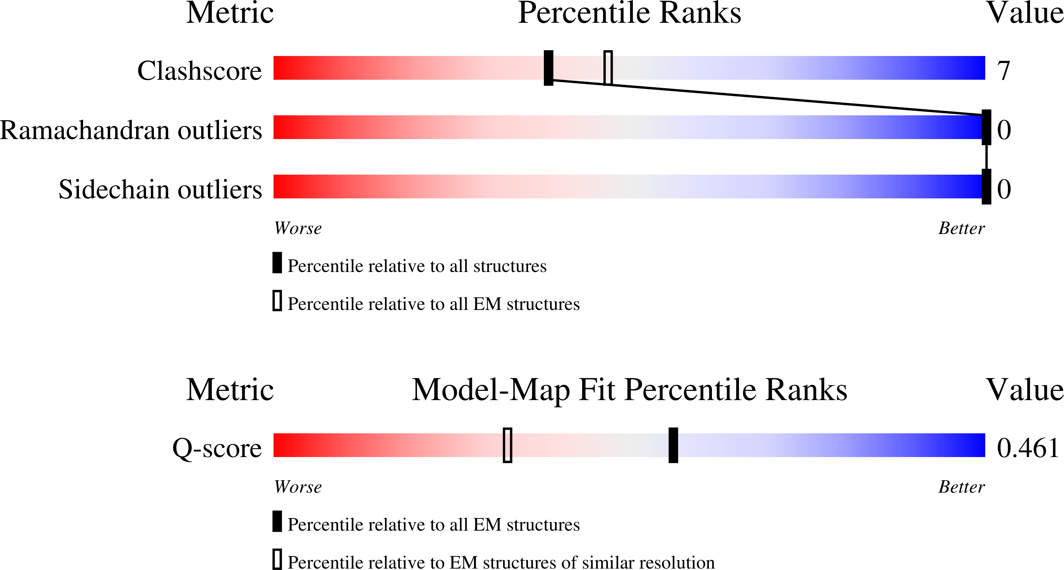
Resolution:
2.97 Å
Aggregation State:
PARTICLE
Reconstruction Method:
SINGLE PARTICLE
Macromolecular Entities
Structures with similar UniProt ID
Protein Blast
Polymer Type:polypeptide(L)
Molecule:Cullin-5
Gene (Uniprot):CUL5
Chain IDs:E (auth: C)
Chain Length:780
Number of Molecules:1
Biological Source:Homo sapiens
Polymer Type:polypeptide(L)
Molecule:L3A2-1
Chain IDs:D (auth: G)
Chain Length:157
Number of Molecules:1
Biological Source:synthetic construct
Structures with similar UniProt ID
Protein Blast
Polymer Type:polypeptide(L)
Molecule:E3 ubiquitin-protein ligase ARIH2
Gene (Uniprot):ARIH2
Chain IDs:F (auth: H)
Chain Length:493
Number of Molecules:1
Biological Source:Homo sapiens
Structures with similar UniProt ID
Protein Blast
Polymer Type:polypeptide(L)
Molecule:NEDD8
Gene (Uniprot):NEDD8
Chain IDs:A (auth: N)
Chain Length:81
Number of Molecules:1
Biological Source:Homo sapiens
Structures with similar UniProt ID
Protein Blast
Polymer Type:polypeptide(L)
Molecule:RING-box protein 2
Gene (Uniprot):RNF7
Chain IDs:B (auth: R)
Chain Length:113
Number of Molecules:1
Biological Source:Homo sapiens
Structures with similar UniProt ID
Protein Blast
Polymer Type:polypeptide(L)
Molecule:Ubiquitin
Gene (Uniprot):UBC
Chain IDs:C (auth: U)
Chain Length:75
Number of Molecules:1
Biological Source:Homo sapiens
Primary Citation
E2 variants for probing E3 ubiquitin ligase activities.
Proc.Natl.Acad.Sci.USA 123 e2524899122 e2524899122 (2026)
PMID: 41481455 DOI: 10.1073/pnas.2524899122

Abstact

E3 ligases partner with E2 enzymes to regulate vast eukaryotic biology. The hierarchical nature of these pairings, with >600 E3s and ~40 E2s in humans, necessitates that E2s cofunction with numerous different E3s. Here, focusing on E3s in the RING-between-RING (RBR) family and their partner UBE2L3 and UBE2D-family E2s, we report an approach to interrogate selected pathways. We screened phage-displayed libraries of structure-based E2 variants (E2Vs) to discover enzymes with enhanced affinity and specificity toward half of all RBR E3 ligases (ARIH1, ARIH2, ANKIB1, CUL9, HOIL1, HOIP, and RNF14). Collectively, these E2Vs allowed distinguishing actions of different cofunctioning E3s, obtaining high-resolution cryogenic Electron Microscopy (cryo-EM) structures of an RBR E3 in the context of a substrate-bound multiprotein complex, and profiling an endogenous RBR E3 response to an extracellular stimulus. Overall, we anticipate that E2V technology will be a generalizable tool to enable in-depth mechanistic and structural analysis of E3 ligase functions, and mapping their activity states and protein partners in cellular signaling cascades.

Legend

Protein

Chemical

Disease

Primary Citation of related structures
Feedback Form
Name
Email
Institute
Feedback