9SD8 image
Deposition Date 2025-08-12
Release Date 2026-02-11
Last Version Date 2026-02-11
Entry Detail
PDB ID:
9SD8
Title:
Crystal structure of C-terminally truncated human PGGHG in complex with glucose.
Biological Source:
Source Organism(s):
Homo sapiens (Taxon ID: 9606)
Expression System(s):
Method Details:
Experimental Method:
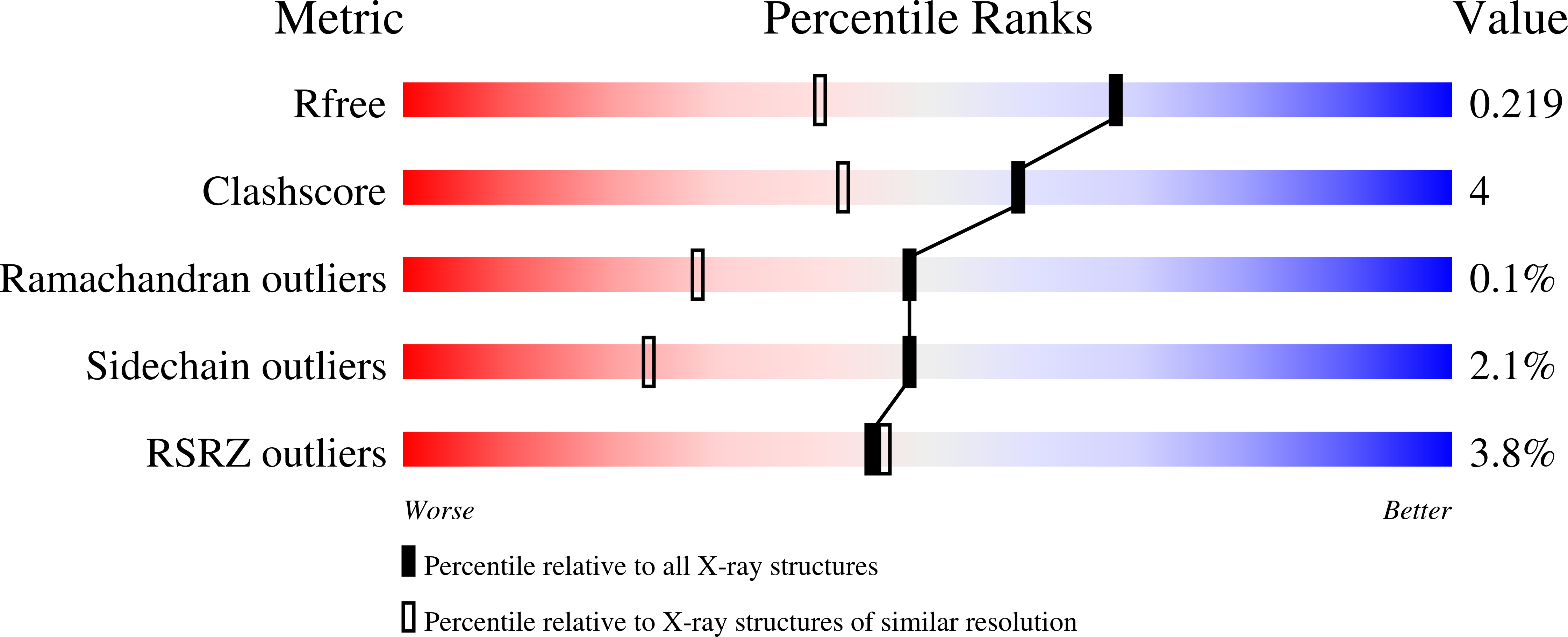
Resolution:
1.61 Å
R-Value Free:
0.20
R-Value Work:
0.17
R-Value Observed:
0.17
Space Group:
P 21 21 21
Macromolecular Entities
Structures with similar UniProt ID
Protein Blast
Polymer Type:polypeptide(L)
Molecule:Protein-glucosylgalactosylhydroxylysine glucosidase
Gene (Uniprot):PGGHG
Mutagens:Deletion of the last 45 C-terminal residues (693-737)
Chain IDs:A, B
Chain Length:693
Number of Molecules:2
Biological Source:Homo sapiens
Primary Citation
The structure of human glucosidase PGGHG reveals a very specific active site accessible through a flat surface for collagen approximation.
Int.J.Biol.Macromol. 345 150556 150556 (2026)
PMID: 41605404 DOI: 10.1016/j.ijbiomac.2026.150556

Abstact

The enzyme glucosylgalactosylhydroxylysine glucosidase (PGGHG) plays a critical role in collagen metabolism by hydrolyzing the 2-O-α-d-glucopyranosyl-O-β-d-galactopyranose, a natural disaccharide found in the glycosylation of hydroxylysine residues in collagen. We report the X-ray crystallographic structure of human PGGHG, revealing the canonical four-domain fold of enzymes from the GH65 family and representing the first structure reported for a mammalian enzyme in this family. A distinctive flat surface adjacent to the catalytic site, shaped by the N-terminal β-sheet and specific conformations of catalytic loops, is unique to PGGHG among GH65 enzymes. Structural complexes with glucose and the substrate analogue kojibiose (KJB), along with site-directed mutagenesis and enzyme assays, identify residues critical for catalysis and hydroxylysine-collagen binding. Docking studies and AlphaFold3-based predictions suggest that the flat surface facilitates the contact between PGGHG and collagen peptides as well as substrate recognition, and support the enzyme's high specificity toward its glucosaccharide substrate. These findings provide structural insight into the selective recognition of glycosylated hydroxylysines and may inform future therapeutic or biotechnological applications targeting collagen metabolism.

Legend

Protein

Chemical

Disease

Primary Citation of related structures
Feedback Form
Name
Email
Institute
Feedback