Deposition Date
2025-08-06
Release Date
2026-01-28
Last Version Date
2026-01-28
Entry Detail
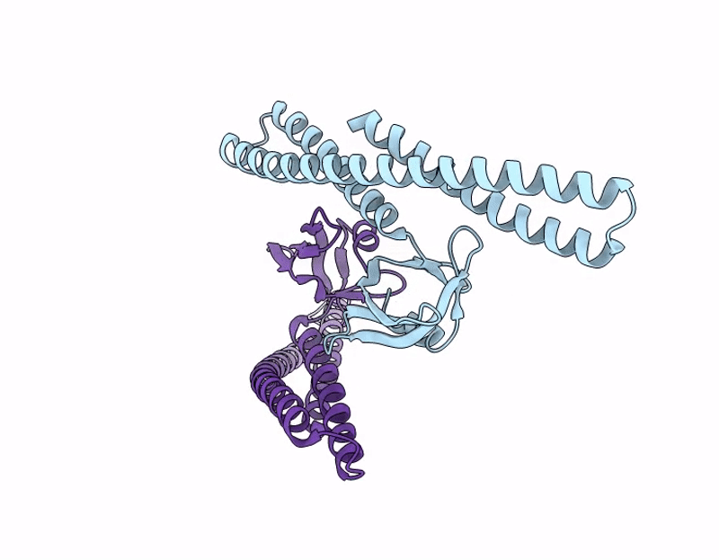
Biological Source:
Source Organism(s):
Saccharolobus islandicus (Taxon ID: 43080)
Expression System(s):
Method Details:
Experimental Method:
Resolution:
4.07 Å
Aggregation State:
FILAMENT
Reconstruction Method:
HELICAL