9RSJ image
Deposition Date 2025-07-01
Release Date 2025-11-26
Last Version Date 2025-11-26
Entry Detail
PDB ID:
9RSJ
Title:
Cryo-EM structure of MATE transporter NorM-VC in complex with doxorubicin
Biological Source:
Source Organism:
Vibrio cholerae (Taxon ID: 666)
synthetic construct (Taxon ID: 32630)
Lama glama (Taxon ID: 9844)
Host Organism:
Method Details:
Experimental Method:
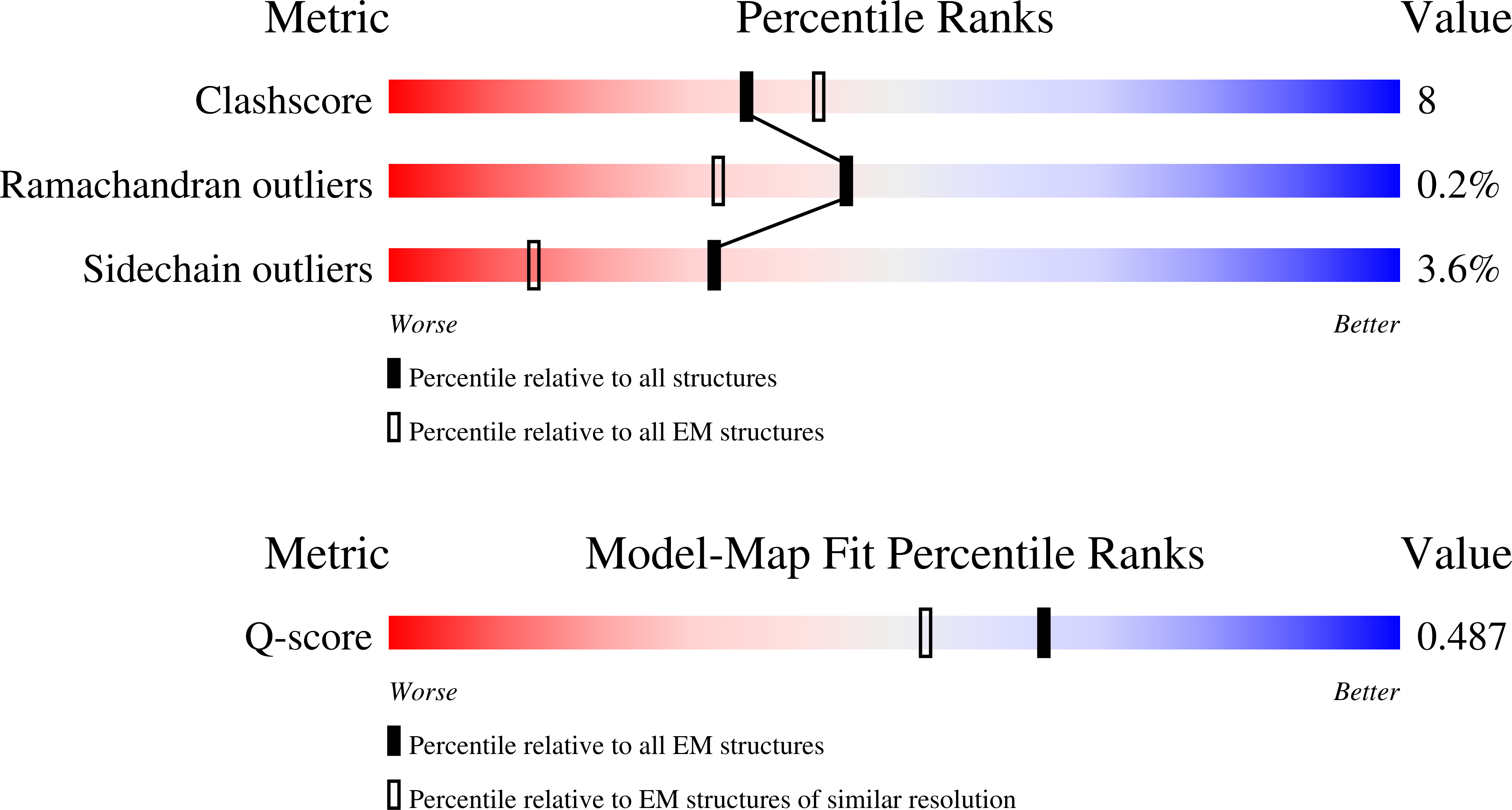
Resolution:
3.10 Å
Aggregation State:
PARTICLE
Reconstruction Method:
SINGLE PARTICLE
Macromolecular Entities
Structures with similar UniProt ID
Protein Blast
Polymer Type:polypeptide(L)
Molecule:Multidrug resistance protein NorM
Gene (Uniprot):VCM66_1481
Chain IDs:A
Chain Length:461
Number of Molecules:1
Biological Source:Vibrio cholerae
Polymer Type:polypeptide(L)
Molecule:NabFab HC
Chain IDs:B (auth: H)
Chain Length:239
Number of Molecules:1
Biological Source:synthetic construct
Polymer Type:polypeptide(L)
Molecule:Anti-Fab nanobody
Chain IDs:E (auth: K)
Chain Length:123
Number of Molecules:1
Biological Source:Lama glama
Polymer Type:polypeptide(L)
Molecule:NabFab LC
Chain IDs:C (auth: L)
Chain Length:215
Number of Molecules:1
Biological Source:synthetic construct
Polymer Type:polypeptide(L)
Molecule:NorM-Nb17_4
Chain IDs:D (auth: N)
Chain Length:132
Number of Molecules:1
Biological Source:synthetic construct
Ligand Molecules
Primary Citation
Doxorubicin recognition and transport by the MATE multidrug transporter NorM from Vibrio cholerae.
J.Mol.Biol. ? 169549 169549 (2025)
PMID: 41260293 DOI: 10.1016/j.jmb.2025.169549

Abstact

Multidrug and toxic compound extrusion (MATE) transport proteins contribute to multidrug resistance in human pathogens by extruding various cytotoxic compounds from the cellular interior. Despite their importance across all domains of life, the specificities and mechanisms of substrate transport of these proteins remain poorly understood due to limited structural and functional information. Here, we determined the cryo-electron microscopy structure of NorM from Vibrio cholerae (NorM-VC) in complex with the anthracycline antibiotic doxorubicin, using the NabFab approach. The structure reveals that the doxorubicin-binding pocket is located halfway through the membrane, within the C-lobe of the protein. Functional studies targeting the doxorubicin-interacting residues validated the binding pocket and enabled detailed analysis of the doxorubicin transport reaction. Our findings indicate doxorubicin binding within a multisite binding chamber engaged in a general transport mechanism for a variety of substrates.

Legend

Protein

Chemical

Disease

Primary Citation of related structures
Feedback Form
Name
Email
Institute
Feedback