9RN6 image
Deposition Date 2025-06-19
Release Date 2026-01-21
Last Version Date 2026-01-21
Entry Detail
PDB ID:
9RN6
Keywords:
Title:
Crystal structure of a protein mimic of SARS-CoV-2 spike's HR1 domain in complex with two nanobodies bound to different epitopes
Biological Source:
Expression System(s):
Method Details:
Experimental Method:
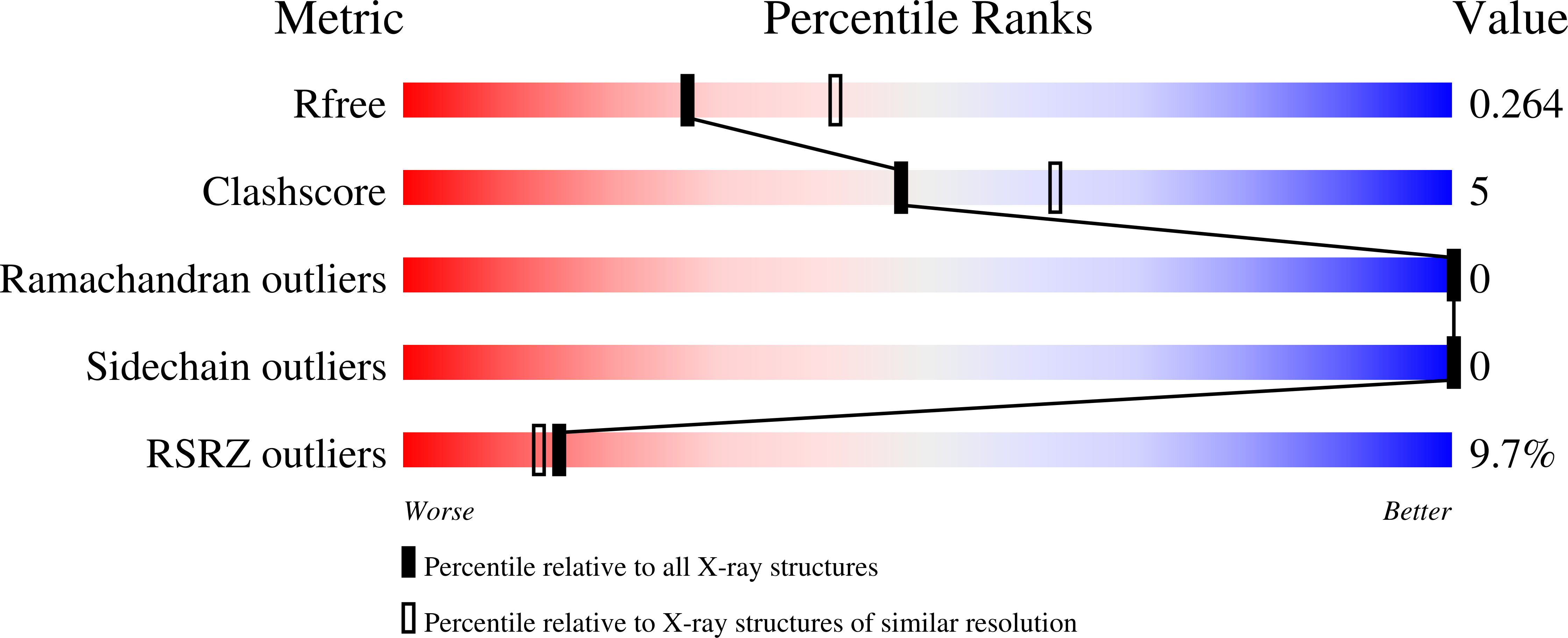
Resolution:
2.40 Å
R-Value Free:
0.26
R-Value Work:
0.24
R-Value Observed:
0.24
Space Group:
P 2 21 21
Macromolecular Entities
Structures with similar UniProt ID
Protein Blast
Polymer Type:polypeptide(L)
Molecule:Spike protein S2',Chimeric protein mimic of SARS-CoV-2 Spike HR1
Gene (Uniprot):S
Chain IDs:A
Chain Length:241
Number of Molecules:1
Biological Source:Severe acute respiratory syndrome coronavirus 2
Polymer Type:polypeptide(L)
Molecule:Nanobody NB278
Chain IDs:B
Chain Length:123
Number of Molecules:1
Biological Source:Vicugna pacos
Polymer Type:polypeptide(L)
Molecule:Nanobody NB184
Chain IDs:C
Chain Length:138
Number of Molecules:1
Biological Source:Vicugna pacos
Ligand Molecules
Primary Citation
Neutralizing nanobodies against SARS-CoV-2 recognizing highly conserved epitopes at the Spike's S2 subunit.
Int.J.Biol.Macromol. 340 150022 150022 (2026)
PMID: 41506506 DOI: 10.1016/j.ijbiomac.2025.150022

Abstact

The formation of a six-helix bundle between the conserved heptad-repeat regions 1 and 2 (HR1 and HR2) in SARS-CoV-2 Spike's S2 subunit is essential for membrane fusion and represents a promising therapeutic target. Previously, we reported recombinant proteins named CoVS-HR1, which mimic the HR1 region and block its interaction with HR2, inhibiting viral fusion. Moreover, they are recognized by plasma antibodies from COVID-19 convalescent patients. In this work, we generated camelid heavy-chain-only antibody fragments (VHHs), also named nanobodies (NBs), against a CoVS-HR1 variant mimicking the full HR1 region. A first generation of selected NBs bound HR1 with high affinity and competed with HR2. Notably, this set of NBs exclusively recognized the C-terminal half of HR1, and two of them showed mild neutralizing activity in cell infection assays. Using a truncated CoVS-HR1 variant (N2C), we selected a second generation of NBs targeting specifically the N-terminal half of HR1. However, these NBs did not demonstrate neutralizing activity, possibly due to their low binding affinities. Several NB epitopes were delineated by hydrogen‑deuterium exchange and mass spectrometry analysis, and the crystal structure of a ternary complex between an HR1-mimetic protein and two NBs was determined, confirming competition with HR2. Intriguingly, we found cooperative binding effects between NBs targeting each half of HR1, but these did not result in detectable inhibitory synergy. These findings demonstrate the existence of neutralizing epitopes in the S2 HR1 region and provide a foundation for future development of enhanced neutralizing NBs focused on specific epitopes using HR1-mimetic proteins.

Legend

Protein

Chemical

Disease

Primary Citation of related structures
Feedback Form
Name
Email
Institute
Feedback