Deposition Date
2025-05-08
Release Date
2025-10-01
Last Version Date
2025-10-08
Method Details:
Experimental Method:
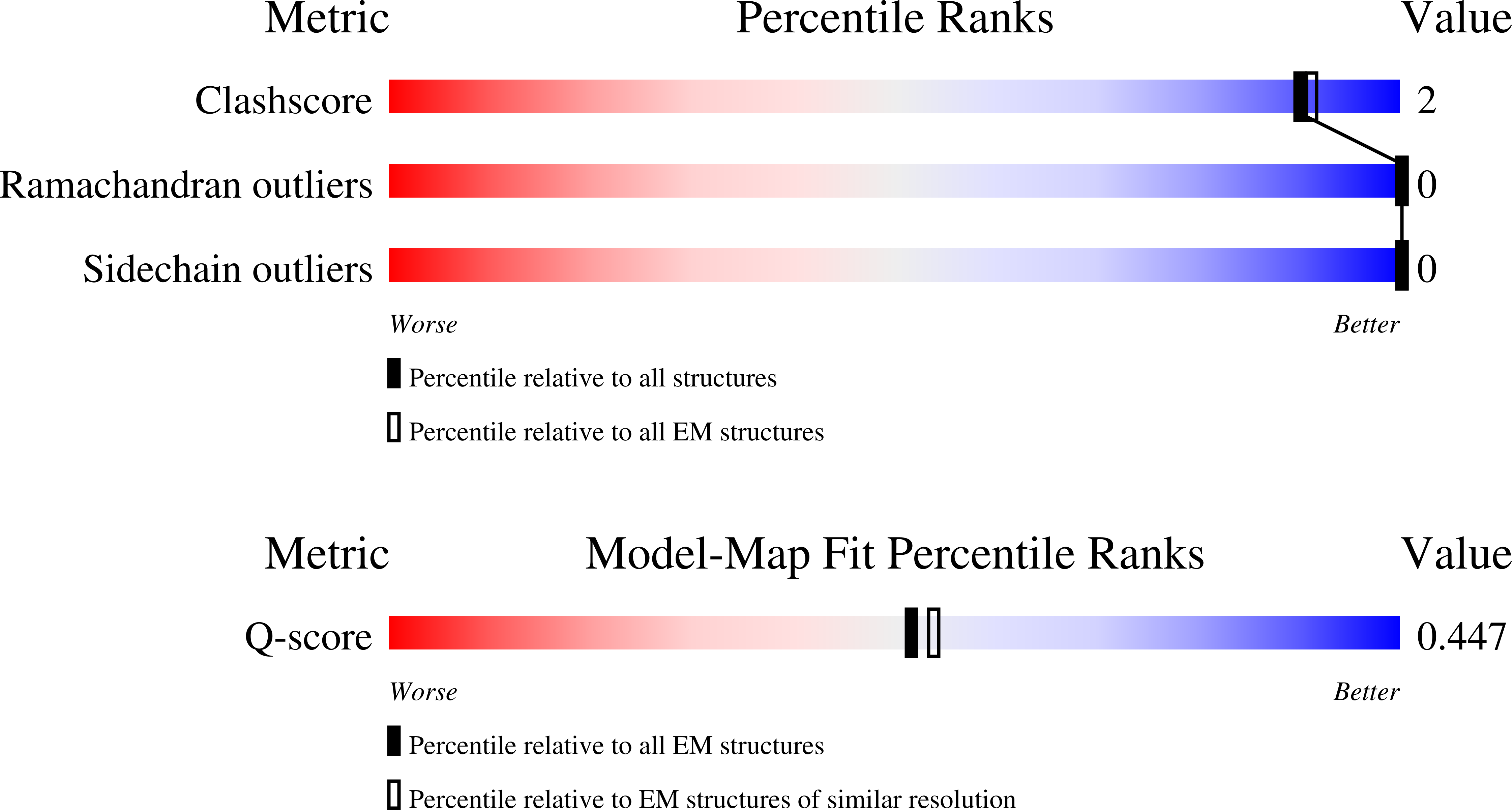
Resolution:
3.50 Å
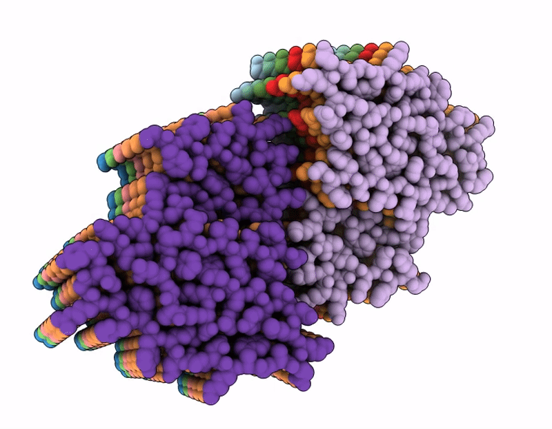
Aggregation State:
HELICAL ARRAY
Reconstruction Method:
HELICAL