9R3Y image
Deposition Date 2025-05-06
Release Date 2025-10-22
Last Version Date 2025-10-22
Entry Detail
PDB ID:
9R3Y
Title:
Solution NMR structure of N-WASP GBD in complex with EspFu R5
Biological Source:
Source Organism:
Homo sapiens (Taxon ID: 9606)
Escherichia coli (Taxon ID: 562)
Host Organism:
Method Details:
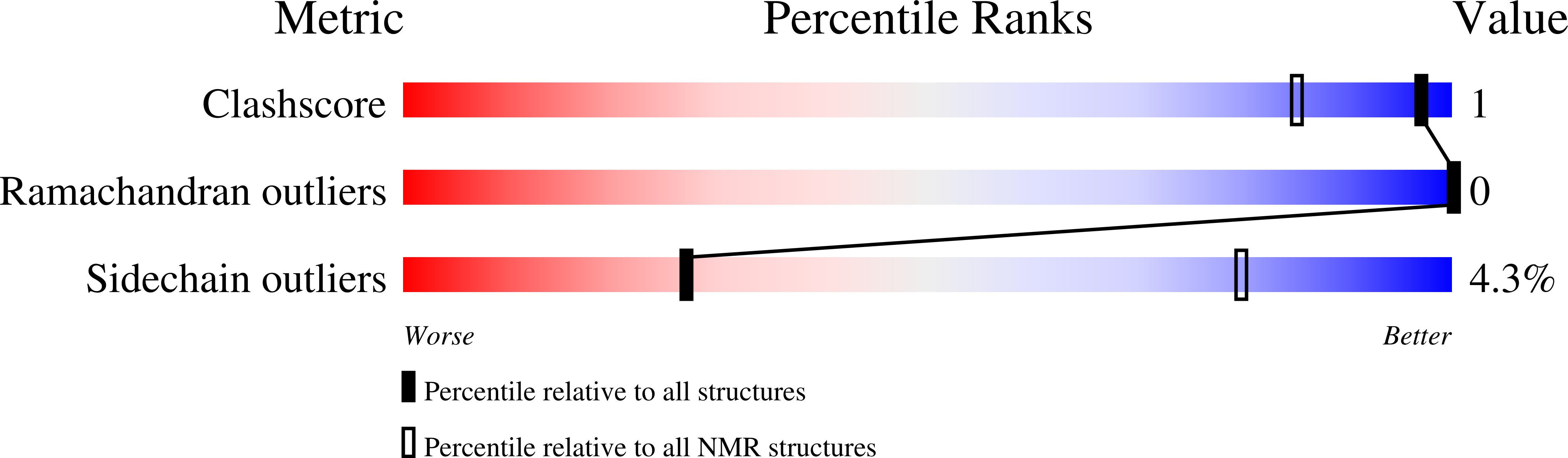
Experimental Method:
Conformers Calculated:
30
Conformers Submitted:
20
Selection Criteria:
structures with the least restraint violations
Macromolecular Entities
Structures with similar UniProt ID
Protein Blast
Polymer Type:polypeptide(L)
Molecule:Actin nucleation-promoting factor WASL
Gene (Uniprot):WASL
Chain IDs:A
Chain Length:73
Number of Molecules:1
Biological Source:Homo sapiens
Structures with similar UniProt ID
Protein Blast
Polymer Type:polypeptide(L)
Molecule:Secreted effector protein EspF(U)
Gene (Uniprot):espF(U)
Chain IDs:B
Chain Length:48
Number of Molecules:1
Biological Source:Escherichia coli
Ligand Molecules
Primary Citation
Intrinsically disordered enteropathogenic E. coli EspF exploits motif mimicry in high-affinity binding to neural Wiskott-Aldrich syndrome protein and sorting nexin 9.
Int.J.Biol.Macromol. 330 148227 148227 (2025)
PMID: 41075884 DOI: 10.1016/j.ijbiomac.2025.148227

Abstact

EspF is an enteropathogenic Escherichia coli (EPEC) effector protein that interferes with intestinal epithelial cell signaling by binding to the Src homology 3 (SH3) domain of sorting nexin 9 (SNX9) and the GTPase-binding domain (GBD) of neural Wiskott-Aldrich syndrome protein (N-WASP) with its C-terminal proline-rich repeats. To understand the molecular basis of these interactions, we characterized the structure, dynamics, and binding thermodynamics of EspF and its target protein domain complexes. We also elaborated on our previous study on EspFU, a homologous effector in enterohemorrhagic E. coli (EHEC), and compared the two effectors. We show that EspF is intrinsically disordered but that NMR chemical shifts expose the pre-structured polyproline II (PPII) helical SH3- and helical GBD-binding motifs. These motifs mimic their cellular counterparts but are fine-tuned to prevail in competitive binding. Factors behind EspF's higher affinity for GBD relative to the cellular ligand are key residue mutations and a C-terminally elongated polar interaction interface. The latter compensates for the lack of an "extended arm", the critical substitution promoting high affinity for GBD in EspFU. With this advantage, EspF outcompetes the autoinhibitory N-WASP C-helix and stimulates actin polymerization. EspF binds SNX9 SH3 with an extended binding interface, residues N-terminal to the RxAPxxP core motif being essential to strong binding. We define the SNX9 SH3-binding epitope as ϕxPxRxAPxxP and propose to re-delineate the EPEC EspF repeat boundaries accordingly. Furthermore, a characteristic 13C secondary chemical shift pattern is recognized as a fingerprint of polyproline II (PPII) helical conformation in the SH3 binding epitope.

Legend

Protein

Chemical

Disease

Primary Citation of related structures
Feedback Form
Name
Email
Institute
Feedback