9R0Q image
Deposition Date 2025-04-24
Release Date 2025-08-06
Last Version Date 2025-08-27
Entry Detail
PDB ID:
9R0Q
Keywords:
Title:
Paraoxonase-1 in complex with terbium(III) and 2-hydroxyquinoline
Biological Source:
Source Organism:
Host Organism:
Method Details:
Experimental Method:
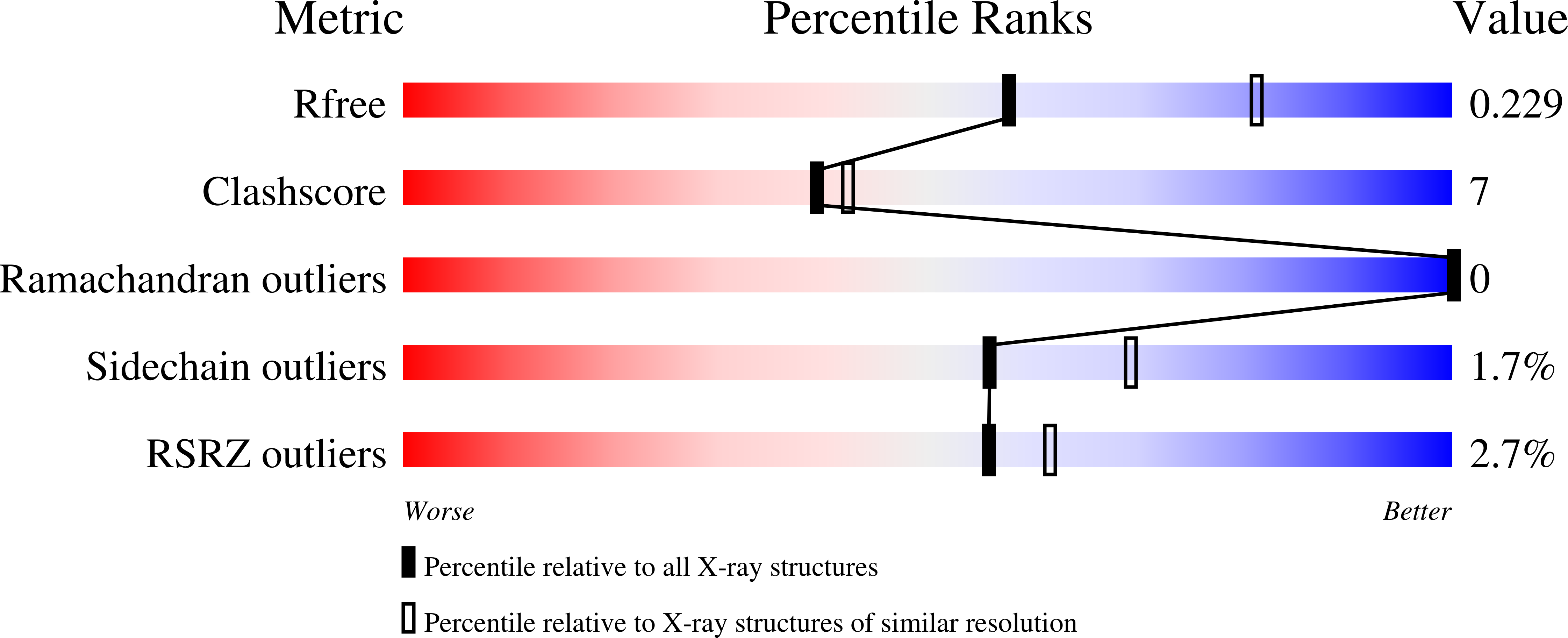
Resolution:
2.35 Å
R-Value Free:
0.22
R-Value Work:
0.19
R-Value Observed:
0.19
Space Group:
P 43 21 2
Macromolecular Entities
Structures with similar UniProt ID
Protein Blast
Polymer Type:polypeptide(L)
Molecule:Serum paraoxonase/arylesterase 1
Gene (Uniprot):PON1
Chain IDs:A
Chain Length:360
Number of Molecules:1
Biological Source:Oryctolagus cuniculus
Primary Citation
Intramolecular sensitization and structure of a Tb 3+ /2-hydroxyquinoline conjugate in the paraoxonase 1 active site.
Dalton Trans 54 12471 12481 (2025)
PMID: 40735812 DOI: 10.1039/d5dt01484k

Abstact

Paraoxonase 1 (PON1) is a Ca2+-dependent enzyme involved in oxidative stress processes and is widely studied for its protective roles in various diseases. Intermolecular sensitization of lanthanide ions was implemented by replacing Ca2+ ions from the recombinant PON1 (rePON1) catalytic site in the presence of 2-hydroxyquinoline (2HQ) as an external antenna. Although the replacement of Ca2+ ions with lanthanide ions indicates weaker binding affinity for the coordination of 2HQ in the protein milieu of the rePON1 active site, it results in the formation of a highly emissive supramolecular complex in the case of Tb3+ ions. The architecture of the ternary rePON1 : Tb3+ : 2HQ conjugate, which allows efficient terbium sensitization and its specific long-wavelength metal phosphorescence emission, was resolved by X-ray crystallography. These findings could establish a non-catalytic quantification strategy for PON1 and provide additional structural insights into lanthanide substitution in this Ca2+-dependent enzyme.

Legend

Protein

Chemical

Disease

Primary Citation of related structures