9QGL image
Deposition Date 2025-03-14
Release Date 2025-10-29
Last Version Date 2025-11-05
Entry Detail
PDB ID:
9QGL
Title:
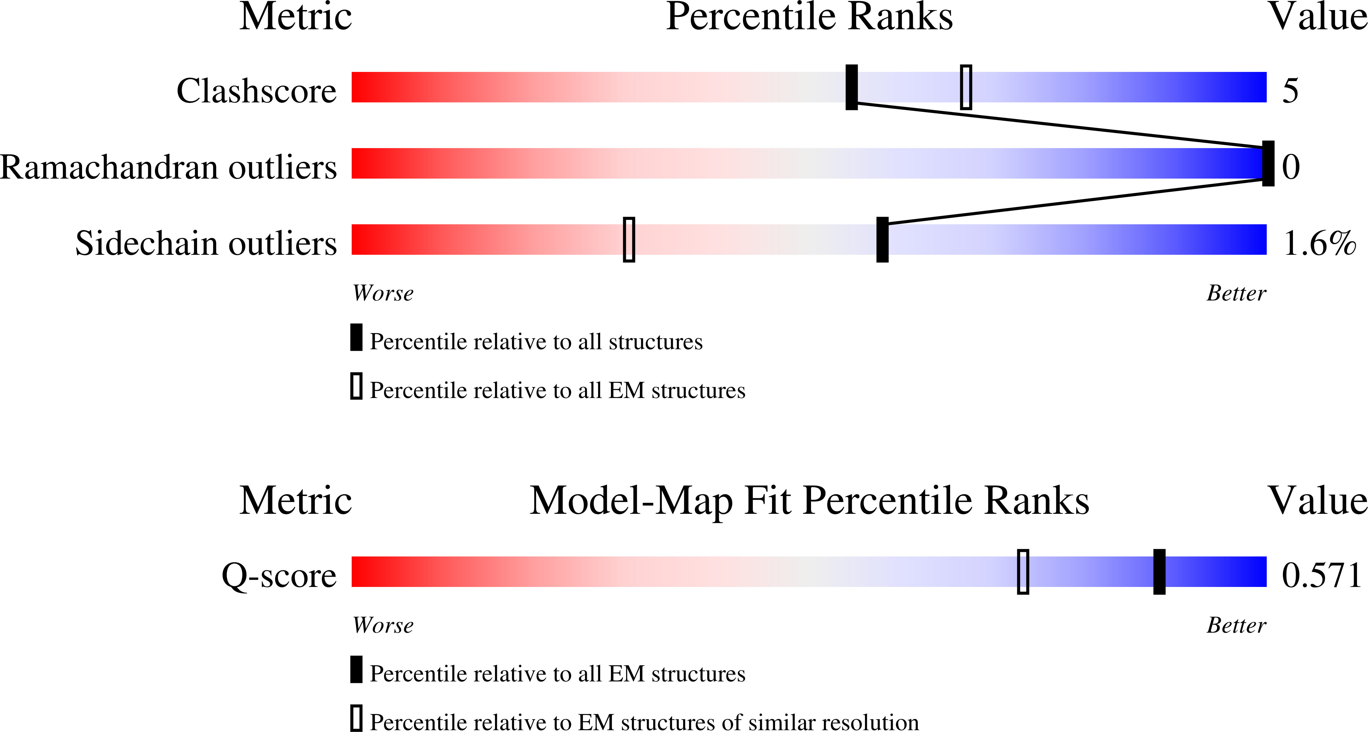
Cryo-EM structure of the PlPVC1 baseplate, 6-fold symmetrized (C6), in extended state
Biological Source:
Source Organism:
Host Organism:
Method Details:
Experimental Method:
Resolution:
2.68 Å
Aggregation State:
PARTICLE
Reconstruction Method:
SINGLE PARTICLE
Macromolecular Entities
Structures with similar UniProt ID
Protein Blast
Polymer Type:polypeptide(L)
Molecule:Phage tail protein
Gene (Uniprot):GPY51_18090
Chain IDs:A (auth: 1A), B (auth: 1B), C (auth: 1C), D (auth: 1D), E (auth: 1E), F (auth: 1F)
Chain Length:149
Number of Molecules:6
Biological Source:Photorhabdus luminescens
Structures with similar UniProt ID
Protein Blast
Polymer Type:polypeptide(L)
Molecule:Phage tail sheath family protein
Gene (Uniprot):GPY51_18095
Chain IDs:G (auth: 2A), H (auth: 2B), I (auth: 2C), J (auth: 2D), K (auth: 2E), L (auth: 2F)
Chain Length:354
Number of Molecules:6
Biological Source:Photorhabdus luminescens
Structures with similar UniProt ID
Protein Blast
Polymer Type:polypeptide(L)
Molecule:Phage tail sheath family protein
Gene (Uniprot):GPY51_18100
Chain IDs:QA (auth: 3A), RA (auth: 3B), SA (auth: 3C), TA (auth: 3D), UA (auth: 3E), VA (auth: 3F)
Chain Length:466
Number of Molecules:6
Biological Source:Photorhabdus luminescens
Structures with similar UniProt ID
Protein Blast
Polymer Type:polypeptide(L)
Molecule:Phage tail sheath family protein
Gene (Uniprot):GPY51_18105
Chain IDs:WA (auth: 4A), XA (auth: 4B), YA (auth: 4C), ZA (auth: 4D), AB (auth: 4E), BB (auth: 4F)
Chain Length:392
Number of Molecules:6
Biological Source:Photorhabdus luminescens
Structures with similar UniProt ID
Protein Blast
Polymer Type:polypeptide(L)
Molecule:Phage tail protein
Gene (Uniprot):C6H66_14800
Chain IDs:M (auth: 5A), N (auth: 5B), O (auth: 5C), P (auth: 5D), Q (auth: 5E), R (auth: 5F)
Chain Length:152
Number of Molecules:6
Biological Source:Photorhabdus luminescens
Structures with similar UniProt ID
Protein Blast
Polymer Type:polypeptide(L)
Molecule:Contractile injection system tube protein N-terminal domain-containing protein
Gene (Uniprot):C6H66_14790
Chain IDs:S (auth: 7A), T (auth: 7B), U (auth: 7C), V (auth: 7D), W (auth: 7E), X (auth: 7F)
Chain Length:227
Number of Molecules:6
Biological Source:Photorhabdus luminescens
Structures with similar UniProt ID
Protein Blast
Polymer Type:polypeptide(L)
Molecule:Photorhabdus luminescens subsp. laumondii TTO1 complete genome segment 6/17
Gene (Uniprot):plu1722
Chain IDs:Y (auth: 9A), Z (auth: 9B), AA (auth: 9C), BA (auth: 9D), CA (auth: 9E), DA (auth: 9F)
Chain Length:239
Number of Molecules:6
Biological Source:Photorhabdus luminescens
Structures with similar UniProt ID
Protein Blast
Polymer Type:polypeptide(L)
Molecule:Photorhabdus luminescens subsp. laumondii TTO1 complete genome segment 6/17
Gene (Uniprot):plu1720
Chain IDs:EA (auth: A), FA (auth: B), GA (auth: C), HA (auth: D), IA (auth: E), JA (auth: F)
Chain Length:905
Number of Molecules:6
Biological Source:Photorhabdus luminescens
Structures with similar UniProt ID
Protein Blast
Polymer Type:polypeptide(L)
Molecule:Baseplate protein J-like domain-containing protein
Gene (Uniprot):GPY51_18145
Chain IDs:KA (auth: a), LA (auth: b), MA (auth: c), NA (auth: d), OA (auth: e), PA (auth: f)
Chain Length:905
Number of Molecules:6
Biological Source:Photorhabdus luminescens
Ligand Molecules
Primary Citation
Structural characterization of an extracellular contractile injection system from Photorhabdus luminescens in extended and contracted states.
Nat Commun 16 9327 9327 (2025)
PMID: 41125615 DOI: 10.1038/s41467-025-64377-z

Abstact

Contractile injection systems (CISs) are phage tail-like nanosyringes that mediate bacterial interactions by puncturing target cell membranes. Within these systems, Photorhabdus Virulence Cassettes (PVCs) can translocate toxins across eukaryotic target cell membranes and have been engineered to deliver diverse protein cargoes into non-natively-targeted organisms. Despite the structural insights into several CISs, including one PVC from P. asymbiotica, information on PVCs from other species and details on the contraction mechanism remain limited. Here, we present the single-particle cryo-electron microscopy structure of PlPVC1, a PVC from the nematode symbiont and insect pathogen Photorhabdus luminescens DJC, in both extended and contracted states. This particle displays distinct structural features that differ from other CISs, such as a cage surrounding the central spike, a larger sheath adaptor, and a plug exposed to the tube lumen. Moreover, we present the structures of the PlPVC1 fiber and the baseplate of the contracted particle, yielding insight into the contraction mechanism. This study provides structural details of the extended and contracted states of the PlPVC1 particle and supports the model in which contraction is triggered. Furthermore, it facilitates the comparison of PlPVC1 with other CISs and expands the scope of engineering opportunities for future biomedical and biotechnological applications.

Legend

Protein

Chemical

Disease

Primary Citation of related structures
Feedback Form
Name
Email
Institute
Feedback