9QCW image
Deposition Date 2025-03-05
Release Date 2025-07-16
Last Version Date 2025-08-06
Entry Detail
PDB ID:
9QCW
Keywords:
Title:
Crystal structure of Rhizobium etli L-asparaginase ReAV K51A mutant
Biological Source:
Source Organism:
Rhizobium etli (Taxon ID: 29449)
Host Organism:
Method Details:
Experimental Method:
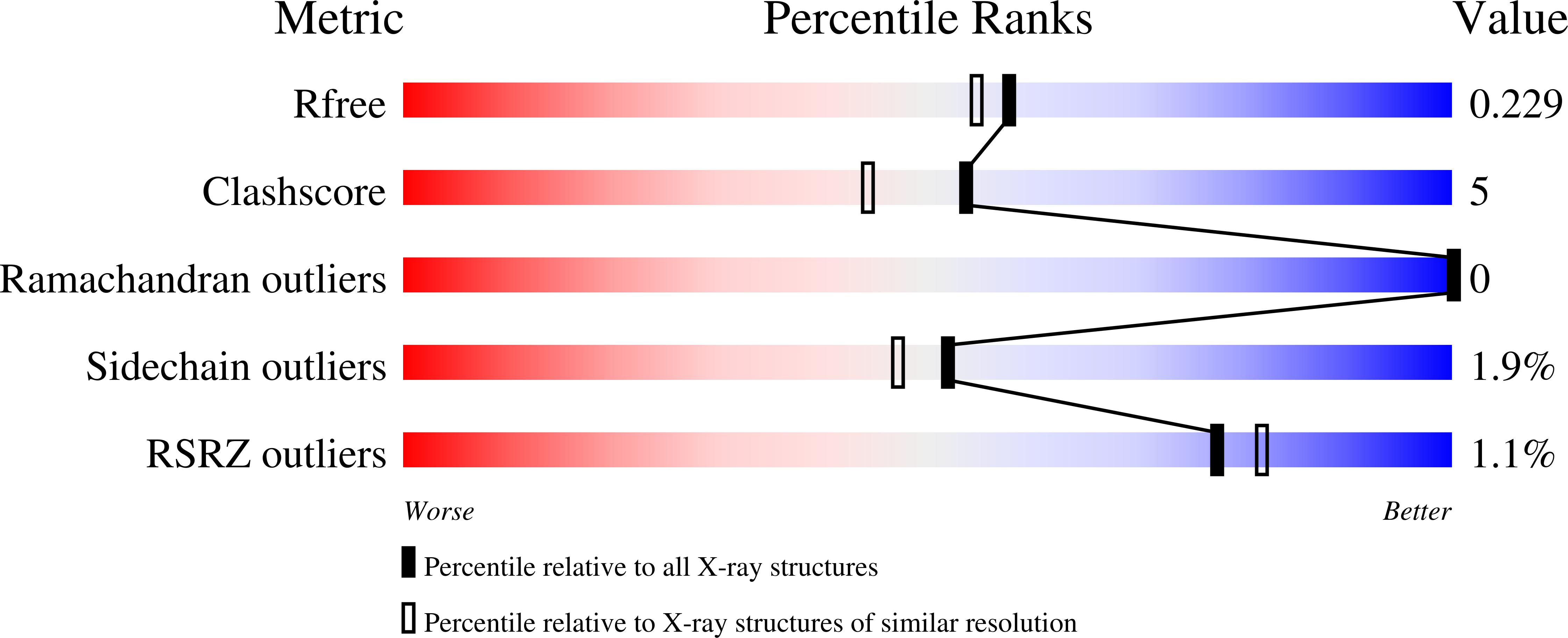
Resolution:
1.95 Å
R-Value Free:
0.22
R-Value Work:
0.18
R-Value Observed:
0.18
Space Group:
P 21 21 21
Macromolecular Entities
Structures with similar UniProt ID
Protein Blast
Polymer Type:polypeptide(L)
Molecule:L-asparaginase II
Gene (Uniprot):ansA
Mutagens:K51A, H238Y
Chain IDs:A, B
Chain Length:373
Number of Molecules:2
Biological Source:Rhizobium etli
Primary Citation

Abstact

The ReAV enzyme from Rhizobium etli, a representative of Class 3 L-asparaginases, is sequentially and structurally different from other known L-asparaginases. This distinctiveness makes ReAV a candidate for novel antileukemic therapies. ReAV is a homodimeric protein, with each subunit containing a highly specific zinc-binding site created by two cysteines, a lysine, and a water molecule. Two Ser-Lys tandems (Ser48-Lys51, Ser80-Lys263) are located in the close proximity of the metal binding site, with Ser48 hypothesized to be the catalytic nucleophile. To further investigate the catalytic process of ReAV, site-directed mutagenesis was employed to introduce alanine substitutions at residues from the Ser-Lys tandems and at Arg47, located near the Ser48-Lys51 tandem. These mutational studies, along with enzymatic assays and X-ray structure determinations, demonstrated that substitution of each of these highly conserved residues abolished the catalytic activity, confirming their essential role in enzyme mechanism.

Legend

Protein

Chemical

Disease

Primary Citation of related structures