9Q8I image
Deposition Date 2025-02-24
Release Date 2025-11-05
Last Version Date 2025-12-03
Entry Detail
PDB ID:
9Q8I
Title:
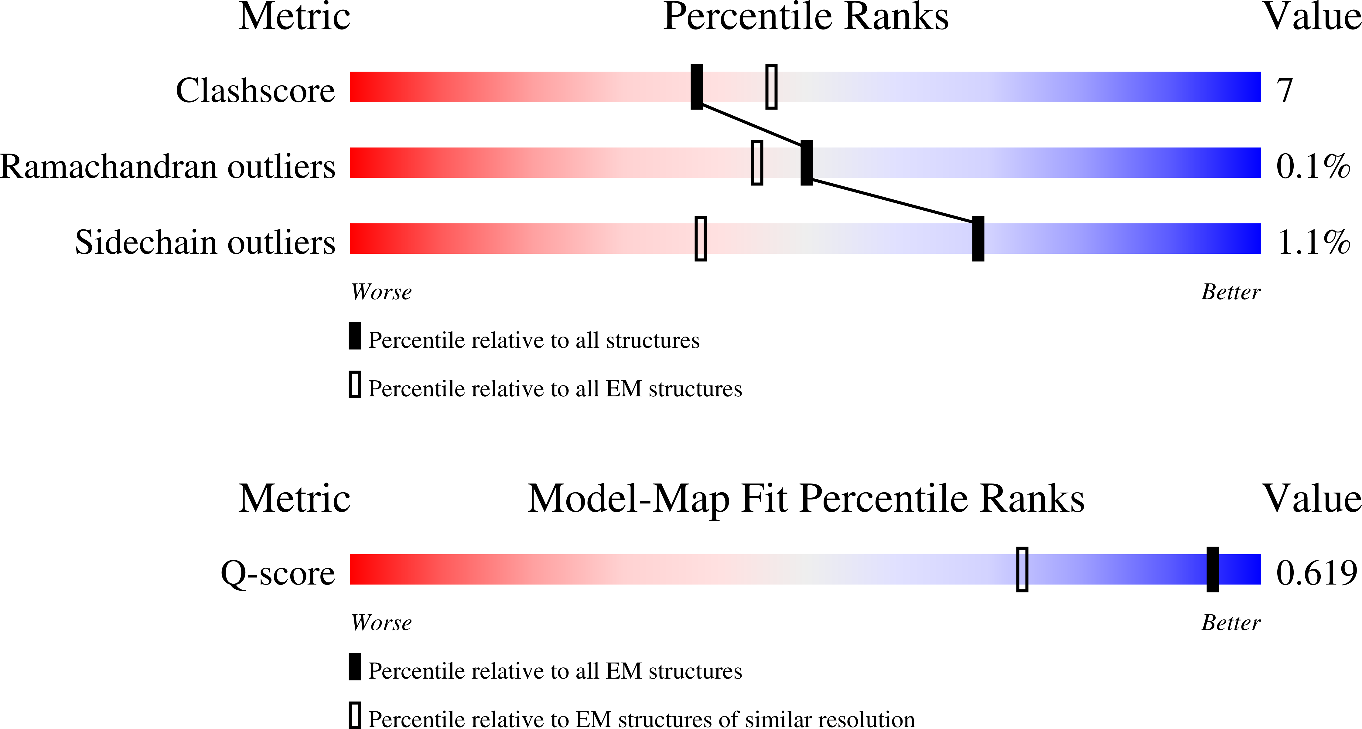
Cryo-EM structure of E. coli complex I variant V96P/N142M (NuoE)
Biological Source:
Source Organism:
Escherichia coli (Taxon ID: 562)
Method Details:
Experimental Method:
Resolution:
2.00 Å
Aggregation State:
PARTICLE
Reconstruction Method:
SINGLE PARTICLE
Macromolecular Entities
Structures with similar UniProt ID
Protein Blast
Polymer Type:polypeptide(L)
Molecule:NADH-quinone oxidoreductase subunit A
Gene (Uniprot):nuoA
Chain IDs:A
Chain Length:147
Number of Molecules:1
Biological Source:Escherichia coli
Structures with similar UniProt ID
Protein Blast
Polymer Type:polypeptide(L)
Molecule:NADH-quinone oxidoreductase subunit B
Gene (Uniprot):nuoB
Chain IDs:B
Chain Length:220
Number of Molecules:1
Biological Source:Escherichia coli
Structures with similar UniProt ID
Protein Blast
Polymer Type:polypeptide(L)
Molecule:NADH-quinone oxidoreductase subunit C/D
Gene (Uniprot):nuoC
Chain IDs:C
Chain Length:596
Number of Molecules:1
Biological Source:Escherichia coli
Structures with similar UniProt ID
Protein Blast
Polymer Type:polypeptide(L)
Molecule:NADH-quinone oxidoreductase subunit E
Gene (Uniprot):nuoE
Mutagens:V96P, N142M
Chain IDs:D (auth: E)
Chain Length:166
Number of Molecules:1
Biological Source:Escherichia coli
Structures with similar UniProt ID
Protein Blast
Polymer Type:polypeptide(L)
Molecule:NADH-quinone oxidoreductase subunit F
Gene (Uniprot):nuoF
Chain IDs:E (auth: F)
Chain Length:461
Number of Molecules:1
Biological Source:Escherichia coli
Structures with similar UniProt ID
Protein Blast
Polymer Type:polypeptide(L)
Molecule:NADH-quinone oxidoreductase subunit G
Gene (Uniprot):nuoG
Chain IDs:F (auth: G)
Chain Length:908
Number of Molecules:1
Biological Source:Escherichia coli
Structures with similar UniProt ID
Protein Blast
Polymer Type:polypeptide(L)
Molecule:NADH-quinone oxidoreductase subunit H
Gene (Uniprot):nuoH
Chain IDs:G (auth: H)
Chain Length:325
Number of Molecules:1
Biological Source:Escherichia coli
Structures with similar UniProt ID
Protein Blast
Polymer Type:polypeptide(L)
Molecule:NADH-quinone oxidoreductase subunit I
Gene (Uniprot):nuoI
Chain IDs:H (auth: I)
Chain Length:180
Number of Molecules:1
Biological Source:Escherichia coli
Structures with similar UniProt ID
Protein Blast
Polymer Type:polypeptide(L)
Molecule:NADH-quinone oxidoreductase subunit J
Gene (Uniprot):nuoJ
Chain IDs:I (auth: J)
Chain Length:184
Number of Molecules:1
Biological Source:Escherichia coli
Structures with similar UniProt ID
Protein Blast
Polymer Type:polypeptide(L)
Molecule:NADH-quinone oxidoreductase subunit K
Gene (Uniprot):nuoK
Chain IDs:J (auth: K)
Chain Length:100
Number of Molecules:1
Biological Source:Escherichia coli
Structures with similar UniProt ID
Protein Blast
Polymer Type:polypeptide(L)
Molecule:NADH-quinone oxidoreductase subunit L
Gene (Uniprot):nuoL
Chain IDs:K (auth: L)
Chain Length:613
Number of Molecules:1
Biological Source:Escherichia coli
Structures with similar UniProt ID
Protein Blast
Polymer Type:polypeptide(L)
Molecule:NADH-quinone oxidoreductase subunit M
Gene (Uniprot):nuoM
Chain IDs:L (auth: M)
Chain Length:509
Number of Molecules:1
Biological Source:Escherichia coli
Structures with similar UniProt ID
Protein Blast
Polymer Type:polypeptide(L)
Molecule:NADH-quinone oxidoreductase subunit N
Gene (Uniprot):nuoN
Chain IDs:M (auth: N)
Chain Length:485
Number of Molecules:1
Biological Source:Escherichia coli
Primary Citation
Structural changes shifting the redox potential of the outlying cluster N1a in respiratory complex I.
Structure ? ? ? (2025)
PMID: 41265450 DOI: 10.1016/j.str.2025.10.016

Abstact

Energy-converting NADH:ubiquinone oxidoreductase, respiratory complex I, is central to energy metabolism by coupling NADH oxidation and quinone reduction with proton translocation across the membrane. Electrons are transferred from the primary acceptor flavin mononucleotide via a chain of iron-sulfur clusters to quinone. The enigmatic cluster N1a is conserved, but not part of this electron transfer chain. We reported on variants of the complex in which N1a is not detectable by EPR spectroscopy. This was tentatively attributed to the lower redox potential of the variant N1a. However, it remained an open question, whether the variants contain this cluster at all. Here, we determined the structures of these variants by X-ray crystallography and cryogenic-electron microscopy. Cluster N1a is present in all variants and the shift of its redox potential is explained by nearby structural changes. A role of the cluster for the mechanism of the complex is discussed.

Legend

Protein

Chemical

Disease

Primary Citation of related structures
Feedback Form
Name
Email
Institute
Feedback