9PZS image
Deposition Date 2025-08-11
Release Date 2025-12-17
Last Version Date 2026-02-04
Entry Detail
PDB ID:
9PZS
Title:
Native GluN1/GluN2A/GluN2B in complex with 5F11 and 3D2 Fabs (class 1), glycine and glutamate bound state
Biological Source:
Source Organism(s):
Mus musculus (Taxon ID: 10090)
Expression System(s):
Method Details:
Experimental Method:
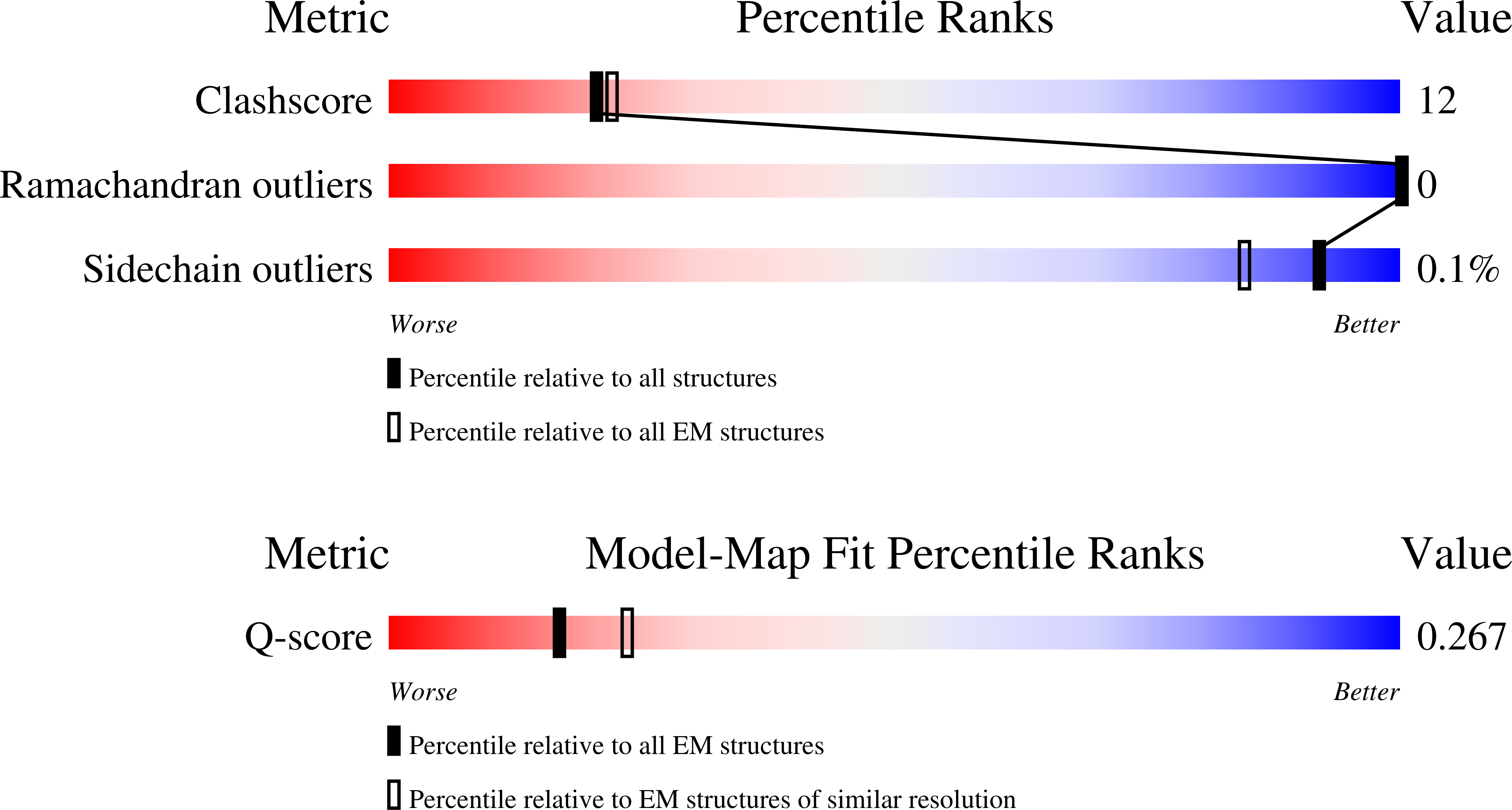
Resolution:
4.20 Å
Aggregation State:
PARTICLE
Reconstruction Method:
SINGLE PARTICLE
Macromolecular Entities
Structures with similar UniProt ID
Protein Blast
Polymer Type:polypeptide(L)
Molecule:Glutamate receptor ionotropic, NMDA 1
Gene (Uniprot):Grin1
Chain IDs:B (auth: A), F (auth: C)
Chain Length:938
Number of Molecules:2
Biological Source:Mus musculus
Structures with similar UniProt ID
Protein Blast
Polymer Type:polypeptide(L)
Molecule:Glutamate receptor ionotropic, NMDA 2A
Gene (Uniprot):Grin2a
Chain IDs:E (auth: B)
Chain Length:1464
Number of Molecules:1
Biological Source:Mus musculus
Structures with similar UniProt ID
Protein Blast
Polymer Type:polypeptide(L)
Molecule:Glutamate receptor ionotropic, NMDA 2B
Gene (Uniprot):Grin2b
Chain IDs:A (auth: D)
Chain Length:1482
Number of Molecules:1
Biological Source:Mus musculus
Polymer Type:polypeptide(L)
Molecule:Heavy chain of GluN1-specific monoclonal Fab fragment, termed 5F11
Chain IDs:C (auth: F), I (auth: H)
Chain Length:277
Number of Molecules:2
Biological Source:Mus musculus
Polymer Type:polypeptide(L)
Molecule:Light chain of GluN1-specific monoclonal Fab fragment, termed 5F11
Chain IDs:D (auth: G), J (auth: I)
Chain Length:244
Number of Molecules:2
Biological Source:Mus musculus
Polymer Type:polypeptide(L)
Molecule:Heavy chain of GluN2A-specific monoclonal Fab fragment, termed 3D2
Chain IDs:G (auth: J)
Chain Length:141
Number of Molecules:1
Biological Source:Mus musculus
Polymer Type:polypeptide(L)
Molecule:Light chain of GluN2A-specific monoclonal Fab fragment, termed 3D2
Chain IDs:H (auth: K)
Chain Length:131
Number of Molecules:1
Biological Source:Mus musculus
Ligand Molecules
Primary Citation
Cryo-EM of autoantibody-bound NMDA receptors reveals antigenic hotspots in an active immunization model of anti-NMDAR encephalitis.
Sci Adv 12 eaeb4249 eaeb4249 (2026)
PMID: 41533802 DOI: 10.1126/sciadv.aeb4249

Abstact

Autoantibodies targeting synaptic membrane proteins are associated with autoimmune encephalitis manifested by seizures, psychosis, and memory dysfunction. Anti-N-methyl-d-aspartate receptor (NMDAR) encephalitis, a prototype of these autoimmune synaptic disorders, is unexpectedly common. Unfortunately, how the native repertoire of anti-NMDAR autoantibodies recognizes NMDARs and the precise locations of antigenic epitopes remain poorly understood. Here, we used an active immunization model that closely mimics the human disease to immunize adult mice with intact GluN1/GluN2A receptors, resulting in fulminant autoimmune encephalitis. Serum was collected at 6 weeks postimmunization for single-particle cryo-electron microscopy of GluN1/GluN2A receptors complexed with purified polyclonal anti-NMDAR autoantibody fragments. Native autoantibodies recognized two distinct binding sites on the GluN1 amino-terminal domain, which we confirmed using monoclonal antibodies bound to native NMDARs purified from mouse brain. Structural analysis of autoantibody-bound NMDAR complexes identified antigenic hotspots within the GluN1 amino-terminal domain. These hotspots provide potential targets for therapeutic intervention.

Legend

Protein

Chemical

Disease

Primary Citation of related structures
Feedback Form
Name
Email
Institute
Feedback