9PXF image
Deposition Date 2025-08-05
Release Date 2026-01-28
Last Version Date 2026-01-28
Entry Detail
PDB ID:
9PXF
Keywords:
Title:
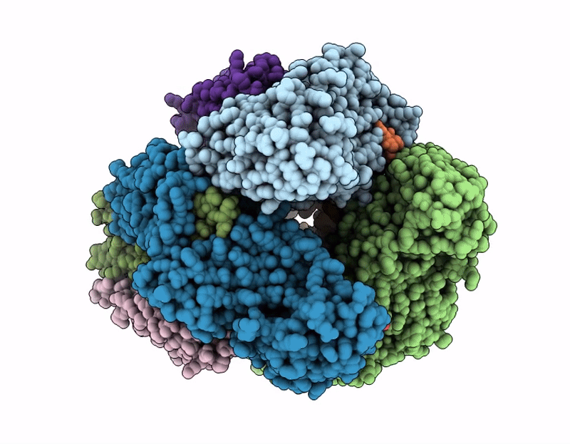
Ammonia monooxygenase in native membranes from N. briensis
Biological Source:
Source Organism(s):
Expression System(s):
Method Details:
Experimental Method:
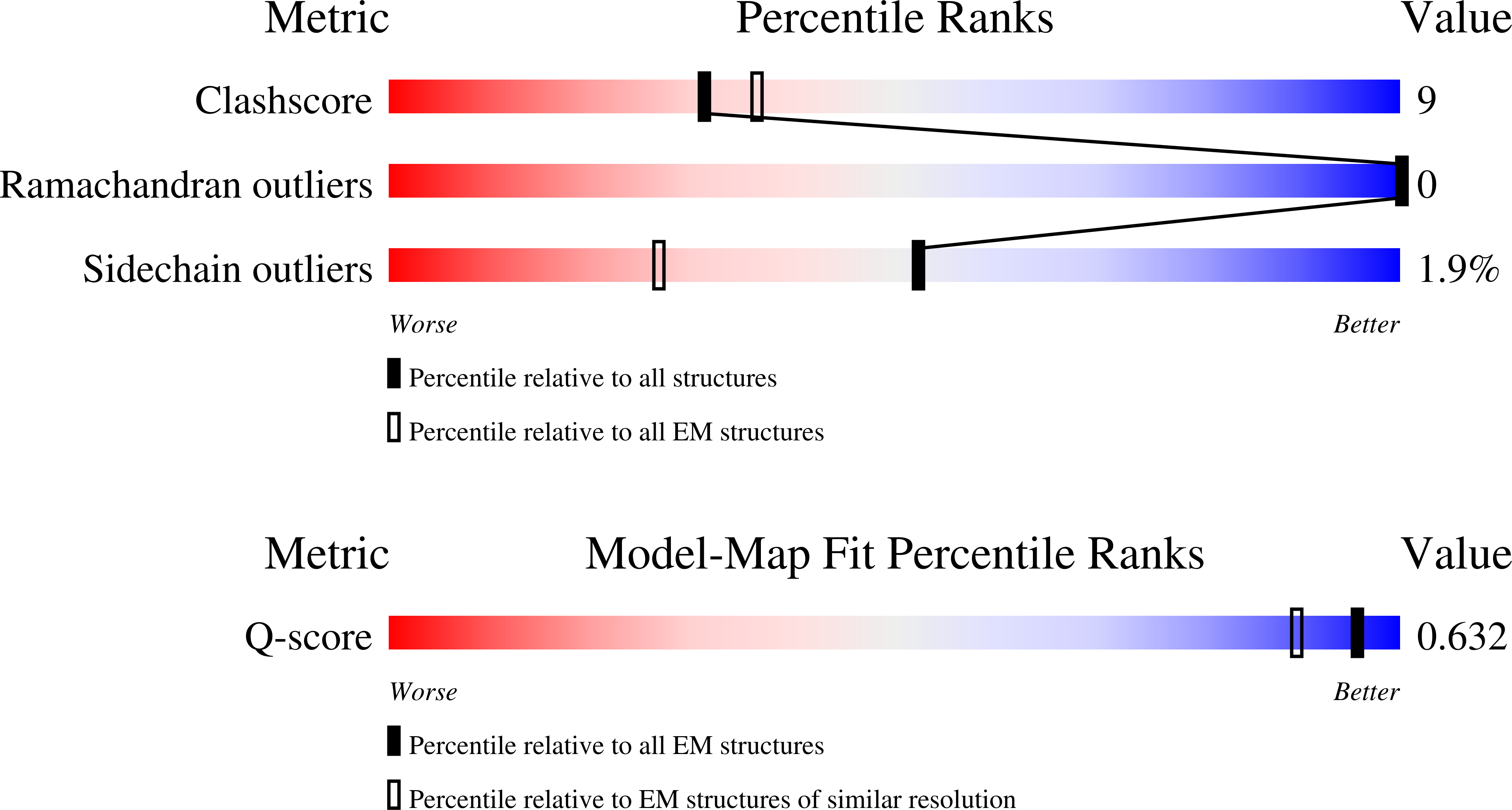
Resolution:
2.58 Å
Aggregation State:
2D ARRAY
Reconstruction Method:
SINGLE PARTICLE
Macromolecular Entities
Structures with similar UniProt ID
Protein Blast
Polymer Type:polypeptide(L)
Molecule:Ammonia monooxygenase subunit B
Gene (Uniprot):SAMN05216386_3024
Chain IDs:C (auth: A), F (auth: B), G (auth: C)
Chain Length:381
Number of Molecules:3
Biological Source:Nitrosospira briensis
Structures with similar UniProt ID
Protein Blast
Polymer Type:polypeptide(L)
Molecule:Ammonia monooxygenase subunit A
Gene (Uniprot):SAMN05216386_3025
Chain IDs:B (auth: D), D (auth: E), E (auth: F)
Chain Length:273
Number of Molecules:3
Biological Source:Nitrosospira briensis
Polymer Type:polypeptide(L)
Molecule:Ammonia monooxygenase subunit C
Chain IDs:A (auth: G), H, I
Chain Length:251
Number of Molecules:3
Biological Source:Nitrosospira briensis
Primary Citation
Simultaneous occupancy of Cu C and Cu D in the ammonia monooxygenase active site.
Chem Sci ? ? ? (2025)
PMID: 41541579 DOI: 10.1039/d5sc08447d

Abstact

Ammonia monooxygenase (AMO), a copper-dependent membrane enzyme, catalyzes the first and rate-limiting step of nitrification: the oxidation of ammonia to hydroxylamine. Despite its central role in the global nitrogen cycle and its biotechnological relevance, structural characterization of AMO has lagged behind that of its homolog, particulate methane monooxygenase (pMMO), due to the slow growth rates of ammonia-oxidizing bacteria and the instability of AMO upon purification. Recent cryoEM studies of Nitrosomonas europaea AMO and Methylococcus capsulatus (Bath) pMMO in native membranes revealed new structural features, including two adjacent copper-binding sites in the transmembrane region, CuC and CuD, believed to constitute the active site. Although multiple structures were determined under various conditions, simultaneous occupancy of CuC and CuD was never observed, leaving their potential functional interplay unresolved. Here we report the 2.6 Å resolution cryoEM structure of AMO from Nitrosospira briensis C-128 in isolated native membranes. This structure reveals the first instance of simultaneous copper occupancy of the CuC and CuD sites, along with occupancy of the periplasmic CuB site. Electron paramagnetic resonance (EPR) spectroscopic data indicate that the CuB site is primarily occupied by Cu(ii), while CuC and CuD are primarily occupied by diamagnetic ions, presumably Cu(i). Notably, a lipid molecule is bound between the CuC and CuD sites, separating them by ∼8.0 Å. The results underscore the importance of studying these enzymes in their native environments across species to resolve conserved and divergent molecular features.

Legend

Protein

Chemical

Disease

Primary Citation of related structures
Feedback Form
Name
Email
Institute
Feedback