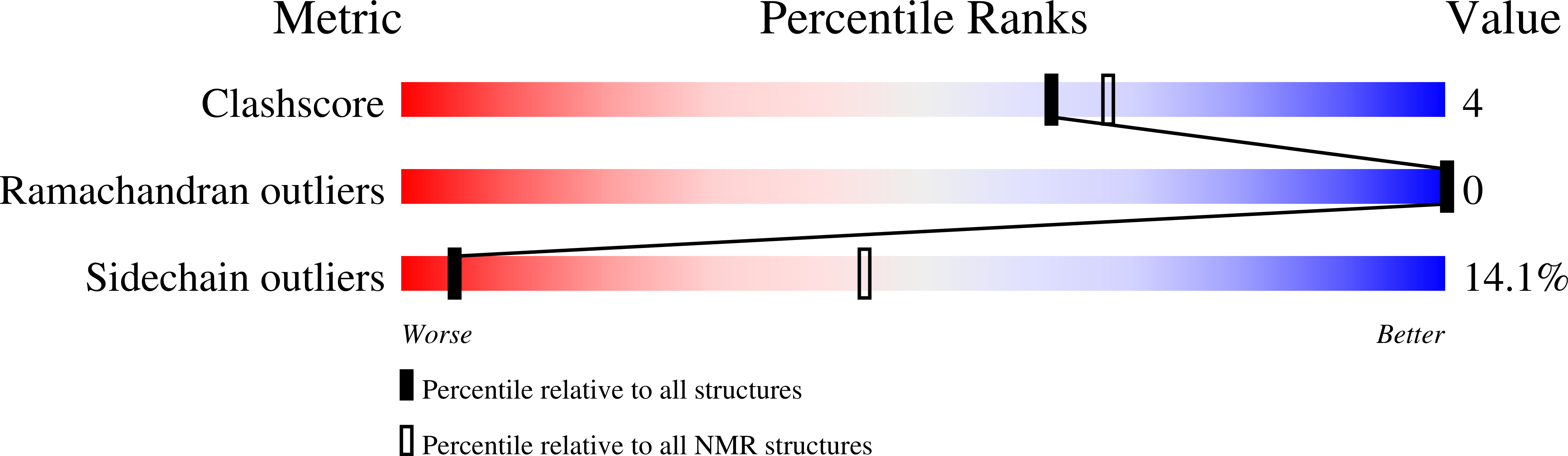
9PQH image
Deposition Date 2025-07-22
Release Date 2026-01-21
Last Version Date 2026-01-21
Entry Detail
PDB ID:
9PQH
Title:
NMR Structure of Ca2+/Calmodulin bound to the GluN1 C0 domain of the NMDA receptor
Biological Source:
Source Organism(s):
Homo sapiens (Taxon ID: 9606)
Expression System(s):
Method Details:
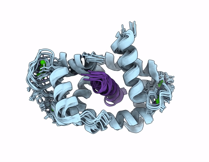
Experimental Method:
Conformers Calculated:
200
Conformers Submitted:
10
Selection Criteria:
structures with the lowest energy
Macromolecular Entities
Structures with similar UniProt ID
Protein Blast
Polymer Type:polypeptide(L)
Molecule:Calmodulin-1
Gene (Uniprot):CALM1
Chain IDs:A
Chain Length:149
Number of Molecules:1
Biological Source:Homo sapiens
Structures with similar UniProt ID
Protein Blast
Polymer Type:polypeptide(L)
Molecule:Glutamate receptor ionotropic, NMDA 1
Gene (Uniprot):GRIN1
Chain IDs:B
Chain Length:938
Number of Molecules:1
Biological Source:Homo sapiens
Ligand Molecules
Primary Citation
Structural Basis and Functional Analysis of NMDA Receptor Regulation by Calmodulin.
J.Biol.Chem. ? 111131 111131 (2026)
PMID: 41513089 DOI: 10.1016/j.jbc.2026.111131

Abstact

The synaptic plasticity mechanisms that are thought to underlie learning and memory require Ca2+ influx mediated by N-methyl-D-aspartate receptors (NMDARs) composed of glycine-binding GluN1 and glutamate-binding GluN2 subunits. Calmodulin (CaM) binding to the cytosolic regions in both GluN1 (residues 841-865, called GluN1-C0) and GluN2A (residues 1004-1023, called GluN2A-C0) may be important for Ca2+-dependent channel desensitization (CDD). Here, we report NMR, ITC and electrophysiological experiments to probe the structure and functional role of Ca2+-bound CaM (Ca2+-CaM) binding to both GluN1 and GluN2A subunits. Our ITC studies show that the GluN1-C0 peptide binds to both the N-lobe and C-lobe of Ca2+-CaM, whereas the GluN2A-C0 peptide binds to only the Ca2+-CaM C-lobe. Our NMR analysis reveals GluN2A residues (W1014 and V1018) interact with exposed hydrophobic residues in the Ca2+-CaM C-lobe. The NMR structure of Ca2+-CaM bound to the GluN1-C0 peptide indicates the two CaM lobes bind to opposite sides of the GluN1-C0 helix (C-lobe contacts M848, F852, A853 and N-lobe contacts A854, V855, W858). The GluN1 mutant F852E and the GluN2A mutant W1014E both perturbed CaM binding in ITC studies, and also diminished electrophysiologically-measured CDD, suggesting CaM interaction with these residues contributes to CDD. We propose a structural mechanism of CDD wherein channel desensitization is caused by the binding of four CaM per NMDAR subunit tetramer.

Legend

Protein

Chemical

Disease

Primary Citation of related structures
Feedback Form
Name
Email
Institute
Feedback