9PFI image
Deposition Date 2025-07-04
Release Date 2025-07-16
Last Version Date 2025-07-16
Entry Detail
PDB ID:
9PFI
Title:
Crystal structure of SARS-CoV-2 Mpro Mutant P132H
Biological Source:
Host Organism:
Method Details:
Experimental Method:
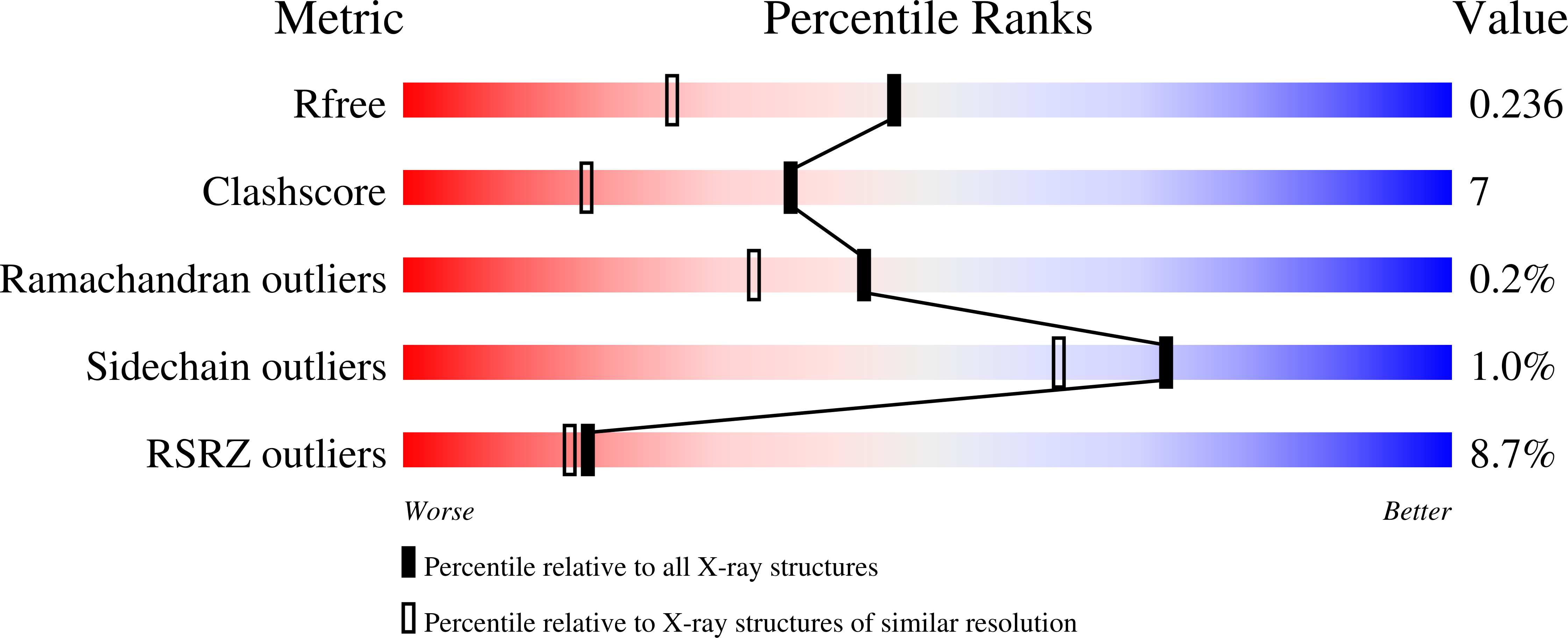
Resolution:
1.81 Å
R-Value Free:
0.23
R-Value Work:
0.18
R-Value Observed:
0.19
Space Group:
P 21 21 21
Macromolecular Entities
Polymer Type:polypeptide(L)
Molecule:3C-like proteinase nsp5
Mutations:P132H
Chain IDs:A, B
Chain Length:306
Number of Molecules:2
Biological Source:Severe acute respiratory syndrome coronavirus 2
Primary Citation
Functional and structural characterization of treatment-emergent nirmatrelvir resistance mutations at low frequencies in the main protease (Mpro) reveals a unique evolutionary route for SARS-CoV-2 to gain resistance.
J Infect Dis ? ? ? (2025)
PMID: 40459233 DOI: 10.1093/infdis/jiaf294

Abstact

BACKGROUND The main protease (Mpro) is one of the most attractive targets for antiviral drug discovery against SARS-CoV-2. Mutations in Mpro have been linked to resistance against nirmatrelvir-ritonavir (NIR-RIT), an important therapy for SARS-CoV-2 infection. This study aimed to identify low-frequency antiviral resistance mutations in Mpro from NIR-RIT-treated patients and to analyze the enzymatic properties, inhibitor susceptibility, and structural features of new Mpro clinical variants. METHODS We screened 1,528 SARS-CoV-2-positive patients from two hospitals and identified 17 who remained positive after treatment. Whole genome sequencing of nasopharyngeal specimens was conducted to identify Mpro clinical variants. The impact of these mutations on Mpro activity and inhibitor susceptibility was investigated using a fluorescent enzymatic biosensor in human cells, along with in vitro thermal stability and structure-based analyses of the Mpro mutants and Mpro-NIR complexes. RESULTS The analysis identified two novel Mpro clinical variants: D48D/L58F/P132H (variant 1) and D48D/L67V/K90R/P132H (variant 2). Our data show that the selected clinical mutations are localized in the Mpro N-terminal domain, are far from the catalytic site, and strongly impact NIR resistance without affecting Mpro activity. Structural analysis and thermal denaturation analyses revealed that these mutations may disrupt the substrate binding site's structure and dynamics, reducing protein stability and potentially impacting substrate binding or dimerization without compromising catalytic activity. CONCLUSIONS Our new Mpro clinical mutations that confer complete resistance to NIR were not identified during previous cell-culture-based studies. More research is needed to explore resistance mechanisms, providing insights into strategies that mitigate resistance and protect therapeutic efficacy.

Legend

Protein

Chemical

Disease

Primary Citation of related structures