9PDI image
Deposition Date 2025-06-30
Release Date 2025-11-19
Last Version Date 2025-11-19
Entry Detail
PDB ID:
9PDI
Title:
Nub1/Fat10-processing human 26S proteasome with Rpt2 at top of spiral staircase and partially unfolded Eos (AAA+ motor locally refined)
Biological Source:
Source Organism:
Homo sapiens (Taxon ID: 9606)
Host Organism:
Method Details:
Experimental Method:
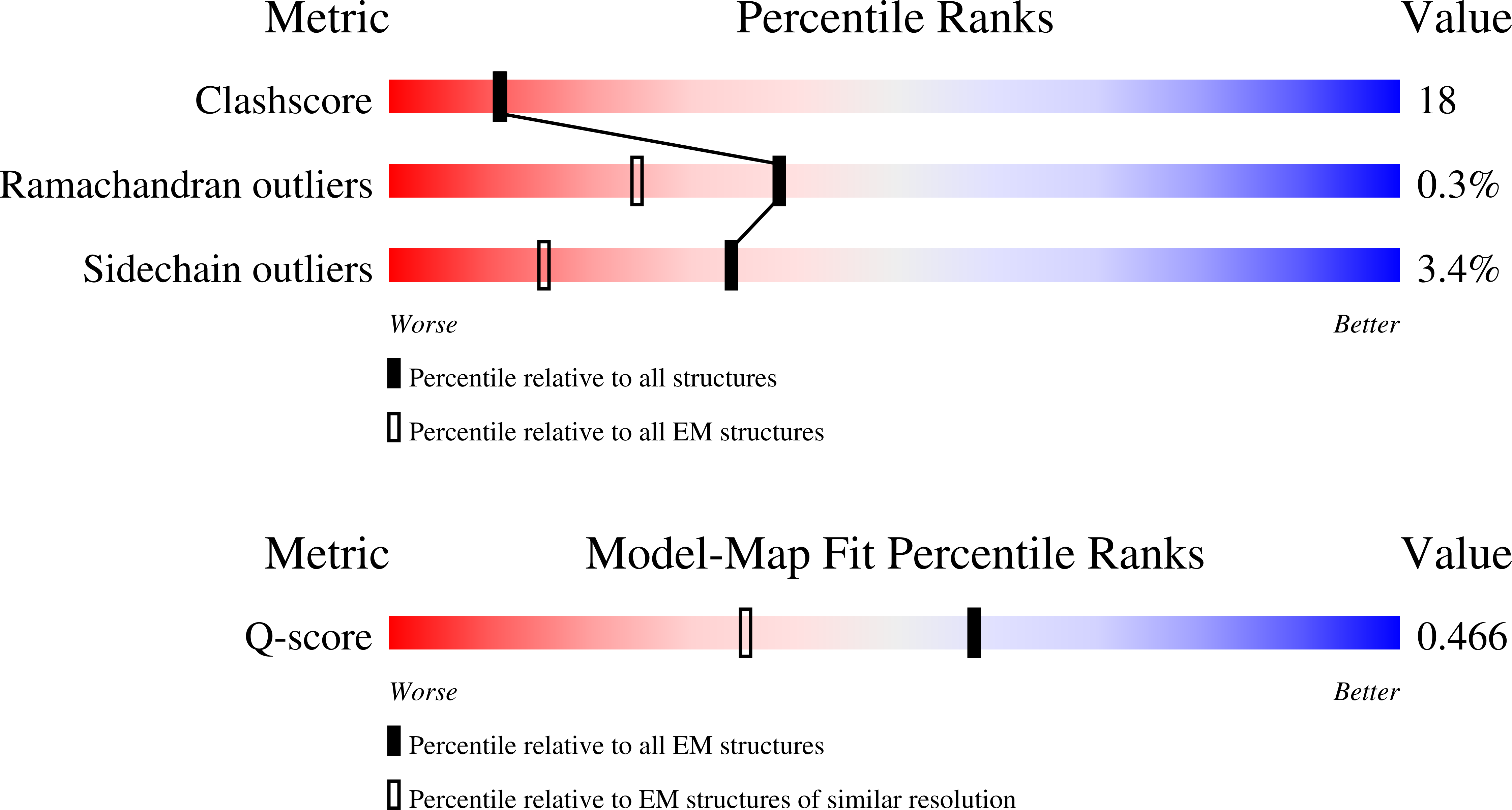
Resolution:
2.98 Å
Aggregation State:
PARTICLE
Reconstruction Method:
SINGLE PARTICLE
Macromolecular Entities
Structures with similar UniProt ID
Protein Blast
Polymer Type:polypeptide(L)
Molecule:26S proteasome regulatory subunit 7
Gene (Uniprot):PSMC2
Chain IDs:L (auth: A)
Chain Length:433
Number of Molecules:1
Biological Source:Homo sapiens
Structures with similar UniProt ID
Protein Blast
Polymer Type:polypeptide(L)
Molecule:26S proteasome regulatory subunit 4
Gene (Uniprot):PSMC1
Chain IDs:M (auth: B)
Chain Length:440
Number of Molecules:1
Biological Source:Homo sapiens
Structures with similar UniProt ID
Protein Blast
Polymer Type:polypeptide(L)
Molecule:26S protease regulatory subunit 8
Gene (Uniprot):PSMC5
Chain IDs:A (auth: C)
Chain Length:424
Number of Molecules:1
Biological Source:Homo sapiens
Structures with similar UniProt ID
Protein Blast
Polymer Type:polypeptide(L)
Molecule:26S proteasome regulatory subunit 6B
Gene (Uniprot):PSMC4
Chain IDs:B (auth: D)
Chain Length:418
Number of Molecules:1
Biological Source:Homo sapiens
Structures with similar UniProt ID
Protein Blast
Polymer Type:polypeptide(L)
Molecule:26S protease regulatory subunit 10B
Gene (Uniprot):PSMC6
Chain IDs:C (auth: E)
Chain Length:389
Number of Molecules:1
Biological Source:Homo sapiens
Structures with similar UniProt ID
Protein Blast
Polymer Type:polypeptide(L)
Molecule:26S proteasome regulatory subunit 6A
Gene (Uniprot):PSMC3
Chain IDs:N (auth: F)
Chain Length:439
Number of Molecules:1
Biological Source:Homo sapiens
Structures with similar UniProt ID
Protein Blast
Polymer Type:polypeptide(L)
Molecule:Proteasome subunit alpha type-6
Gene (Uniprot):PSMA6
Chain IDs:D (auth: G)
Chain Length:246
Number of Molecules:1
Biological Source:Homo sapiens
Structures with similar UniProt ID
Protein Blast
Polymer Type:polypeptide(L)
Molecule:Proteasome subunit alpha type-2
Gene (Uniprot):PSMA2
Chain IDs:E (auth: H)
Chain Length:234
Number of Molecules:1
Biological Source:Homo sapiens
Structures with similar UniProt ID
Protein Blast
Polymer Type:polypeptide(L)
Molecule:Proteasome subunit alpha type-4
Gene (Uniprot):PSMA4
Chain IDs:F (auth: I)
Chain Length:261
Number of Molecules:1
Biological Source:Homo sapiens
Structures with similar UniProt ID
Protein Blast
Polymer Type:polypeptide(L)
Molecule:Proteasome subunit alpha type-7
Gene (Uniprot):PSMA7
Chain IDs:G (auth: J)
Chain Length:248
Number of Molecules:1
Biological Source:Homo sapiens
Structures with similar UniProt ID
Protein Blast
Polymer Type:polypeptide(L)
Molecule:Proteasome subunit alpha type-5
Gene (Uniprot):PSMA5
Chain IDs:H (auth: K)
Chain Length:241
Number of Molecules:1
Biological Source:Homo sapiens
Structures with similar UniProt ID
Protein Blast
Polymer Type:polypeptide(L)
Molecule:Proteasome subunit alpha type-1
Gene (Uniprot):PSMA1
Chain IDs:I (auth: L)
Chain Length:263
Number of Molecules:1
Biological Source:Homo sapiens
Structures with similar UniProt ID
Protein Blast
Polymer Type:polypeptide(L)
Molecule:Proteasome subunit alpha type-3
Gene (Uniprot):PSMA3
Chain IDs:J (auth: M)
Chain Length:255
Number of Molecules:1
Biological Source:Homo sapiens
Structures with similar UniProt ID
Protein Blast
Polymer Type:polypeptide(L)
Molecule:26S proteasome non-ATPase regulatory subunit 14
Gene (Uniprot):PSMD14
Chain IDs:O (auth: c)
Chain Length:424
Number of Molecules:1
Biological Source:Homo sapiens
Polymer Type:polypeptide(L)
Molecule:Substrate polypeptide
Chain IDs:K (auth: v)
Chain Length:22
Number of Molecules:1
Biological Source:Homo sapiens
Primary Citation
Structural landscape of the degrading 26S proteasome reveals conformation-specific binding of TXNL1.
Nat.Struct.Mol.Biol. ? ? ? (2025)
PMID: 41198955 DOI: 10.1038/s41594-025-01695-2

Abstact

The 26S proteasome targets many cellular proteins for degradation during homeostasis and quality control. Proteasome-interacting cofactors modulate these functions and aid in substrate degradation. Here we solve high-resolution structures of the redox active cofactor TXNL1 bound to the human 26S proteasome at saturating and substoichiometric concentrations by time-resolved cryo-electron microscopy (cryo-EM). We identify distinct binding modes of TXNL1 that depend on the proteasome conformation and ATPase motor states. Together with biophysical and biochemical experiments, we show that the resting-state proteasome binds TXNL1 with low affinity and in variable positions on top of the Rpn11 deubiquitinase. In contrast, in the actively degrading proteasome, TXNL1 uses additional interactions for high-affinity binding, whereby its C-terminal tail covers the catalytic groove of Rpn11 and coordinates the active-site Zn2+. Furthermore, these cryo-EM structures of the degrading proteasome capture the ATPase hexamer in several spiral-staircase arrangements that indicate temporally asymmetric hydrolysis and conformational changes in bursts during mechanical substrate unfolding and translocation. Remarkably, we catch the proteasome in the act of unfolding the β-barrel mEos3.2 substrate while the ATPase hexamer is in a particular staircase register. Our findings advance current models for protein translocation through hexameric AAA+ motors and reveal how the proteasome uses its distinct conformational states to coordinate cofactor binding and substrate processing.

Legend

Protein

Chemical

Disease

Primary Citation of related structures
Feedback Form
Name
Email
Institute
Feedback