9PCS image
Deposition Date 2025-06-29
Release Date 2025-10-08
Last Version Date 2025-10-08
Entry Detail
PDB ID:
9PCS
Keywords:
Title:
Crystal structure of Dihydrodipicolinate Synthase from Mycobacterium tuberculosis in complex with pyruvate
Biological Source:
Source Organism:
Host Organism:
Method Details:
Experimental Method:
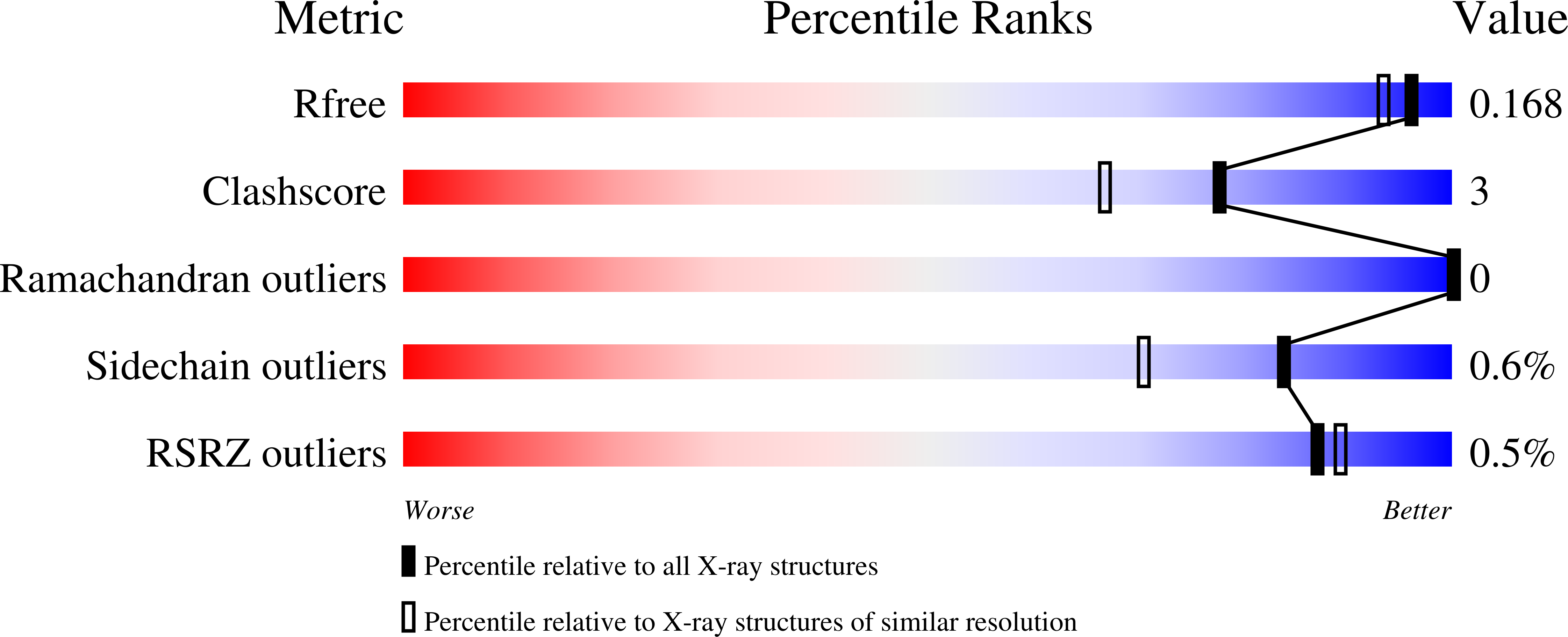
Resolution:
1.50 Å
R-Value Free:
0.16
R-Value Work:
0.15
R-Value Observed:
0.15
Space Group:
P 1 21 1
Macromolecular Entities
Structures with similar UniProt ID
Protein Blast
Polymer Type:polypeptide(L)
Molecule:4-hydroxy-tetrahydrodipicolinate synthase
Gene (Uniprot):dapA
Chain IDs:A, B, C, D, E, F, G, H
Chain Length:300
Number of Molecules:8
Biological Source:Mycobacterium tuberculosis H37Rv
Primary Citation
Crystal structure of dihydrodipicolinate synthase from Mycobacterium tuberculosis in complex with pyruvate and insights into allosteric regulation.
Int.J.Biol.Macromol. 330 147950 147950 (2025)
PMID: 41022234 DOI: 10.1016/j.ijbiomac.2025.147950

Abstact

Tuberculosis is an infectious disease caused by Mycobacterium tuberculosis (Mtb) and is one of the leading causes of death worldwide. This disease is typically treated by combining several antimicrobials for extended periods, which can lead to treatment interruptions by patients and promote the emergence of multidrug-resistant strains, necessitating the use of alternative or second-line drugs. In this perspective, dihydrodipicolinate synthase (DapA) from M. tuberculosis (MtDapA), which catalyzes the aldol condensation between pyruvate and aspartate-semialdehyde (ASA) to produce dihydrodipicolinate, is an essential enzyme in Mtb for the production of l-lysine and meso-diaminopimelate (mDAP). Through crystallographic assays, we have determined the structure of MtDapA in complex with its substrate, pyruvate, covalently bonded through a Schiff base to the catalytic l-lysine at a resolution of 1.5 Å. Through structural analysis, we describe the arrangement of interactions between the active site amino acid residues and pyruvate, providing insight into the binding mode of this molecule. In addition, we performed further biophysical assays, including differential scanning fluorimetry (DSF) and isothermal titration calorimetry (ITC), to obtain insights into the pyruvate affinity and the potential role of l-lysine and mDAP as allosteric regulators of MtDapA. However, in contrast to those observed in other orthologous enzymes, particularly those from Gram-negative bacteria, MtDapA does not have an affinity for l-lysine or mDAP. Consequently, this enzyme is not allosterically regulated by the products of this pathway. The results shown here provide evidence regarding the functioning of the enzyme regulatory mechanism and valuable structural features to aid in the future development of MtDapA inhibitors, which may be further explored in drug discovery campaigns against tuberculosis.

Legend

Protein

Chemical

Disease

Primary Citation of related structures
Feedback Form
Name
Email
Institute
Feedback