9P2R image
Deposition Date 2025-06-12
Release Date 2025-11-12
Last Version Date 2025-11-12
Entry Detail
PDB ID:
9P2R
Title:
Extended, CYR715-bound state of Manduca sexta soluble guanylate cyclase mutant beta C122S
Biological Source:
Source Organism:
Manduca sexta (Taxon ID: 7130)
Method Details:
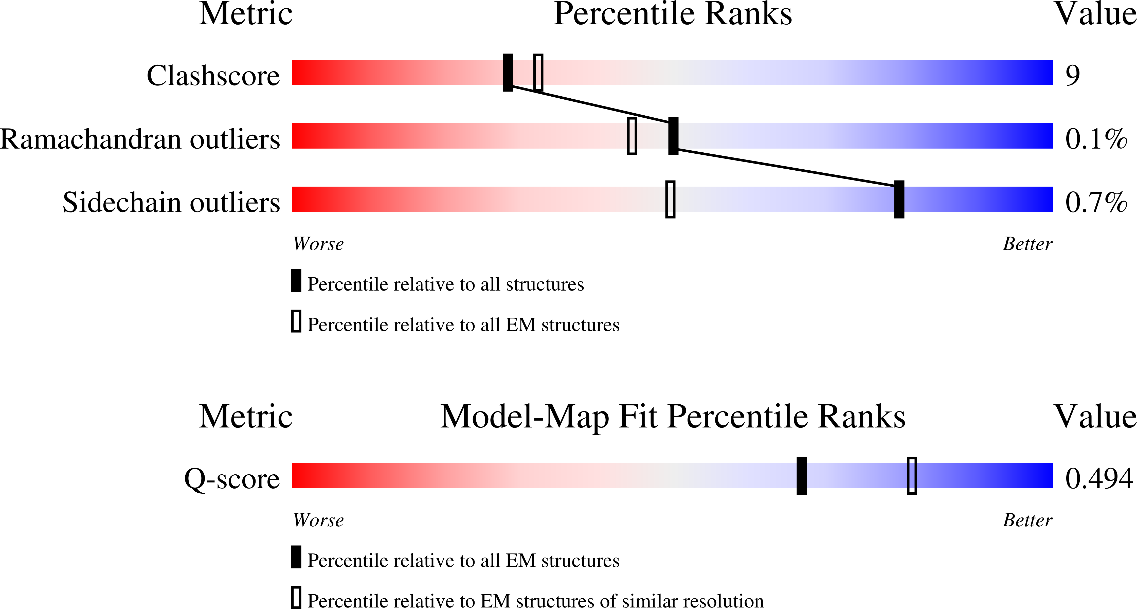
Experimental Method:
Resolution:
3.60 Å
Aggregation State:
PARTICLE
Reconstruction Method:
SINGLE PARTICLE
Macromolecular Entities
Structures with similar UniProt ID
Protein Blast
Polymer Type:polypeptide(L)
Molecule:Soluble guanylyl cyclase alpha-1 subunit
Chain IDs:A
Chain Length:699
Number of Molecules:1
Biological Source:Manduca sexta
Structures with similar UniProt ID
Protein Blast
Polymer Type:polypeptide(L)
Molecule:Guanylate cyclase soluble subunit beta-1
Mutagens:C122S
Chain IDs:B
Chain Length:600
Number of Molecules:1
Biological Source:Manduca sexta
Primary Citation
Molecular Aspects of Soluble Guanylate Cyclase Activation and Stimulator Function.
Biochemistry ? ? ? (2025)
PMID: 41146038 DOI: 10.1021/acs.biochem.5c00424

Abstact

Soluble guanylate cyclases (sGCs) are heme-containing, gas-sensing proteins which catalyze the formation of cGMP from GTP. In humans, sGCs are highly selective sensors of nitric oxide (NO) and play a critical role in NO-based regulation of cardiovascular and pulmonary function. The physiological importance of sGC signaling has led to the development of drugs, known as stimulators and activators, which increase sGC catalytic function. Here we characterize a newly developed stimulator, CYR715, which is a particularly potent stimulator of Manduca sexta (Ms) sGC catalytic function even in the absence of NO, increasing activity of the NO-free enzyme to 45% of full catalytic activity. CYR715 also increased the catalytic activity of Ms sGC βC122A and βC122S variants, with a marked stimulation of the NO-free βC122S variant to 74% of maximum. High-resolution cryo-electron microscopy structures were solved for CYR715 bound to Ms sGC βC122S revealing that CYR715 occupies the same binding site as the characterized sGC stimulators YC-1 and riociguat. Additionally, the core scaffold of CYR715 makes a binding interaction with βC78 while the flexible tail can interact with αR429 or βY7 and E361. Conformational extension of sGC following NO, YC-1, or CYR715 binding was characterized using small-angle X-ray scattering, revealing that while ligand binding results in sGC extension this extension does not directly correlate to observed activity. This suggests that not all conformational extensions of sGC result in increased catalytic activity, and that effective stimulators assist in converting extension into catalytic function.

Legend

Protein

Chemical

Disease

Primary Citation of related structures
Feedback Form
Name
Email
Institute
Feedback