Deposition Date
2025-06-07
Release Date
2025-08-27
Last Version Date
2025-12-17
Entry Detail
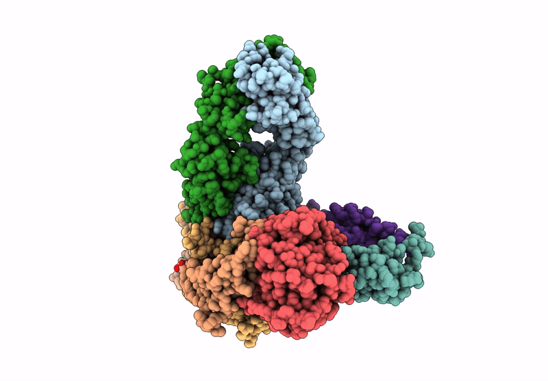
PDB ID:
9P0X
Keywords:
Title:
Nanodisc-embedded human TF/FVIIa/XK1 in complex with 10H10 Fab (nanodisc-subtracted)
Biological Source:
Source Organism(s):
Homo sapiens (Taxon ID: 9606)
Escherichia coli (Taxon ID: 562)
Mus musculus (Taxon ID: 10090)
Escherichia coli (Taxon ID: 562)
Mus musculus (Taxon ID: 10090)
Expression System(s):
Method Details:
Experimental Method:
Resolution:
3.70 Å
Aggregation State:
PARTICLE
Reconstruction Method:
SINGLE PARTICLE