9OMJ image
Deposition Date 2025-05-14
Release Date 2025-10-29
Last Version Date 2025-11-12
Entry Detail
PDB ID:
9OMJ
Keywords:
Title:
WrtF fucosyltransferase - GDP-beta-L-fucose
Biological Source:
Source Organism:
Method Details:
Experimental Method:
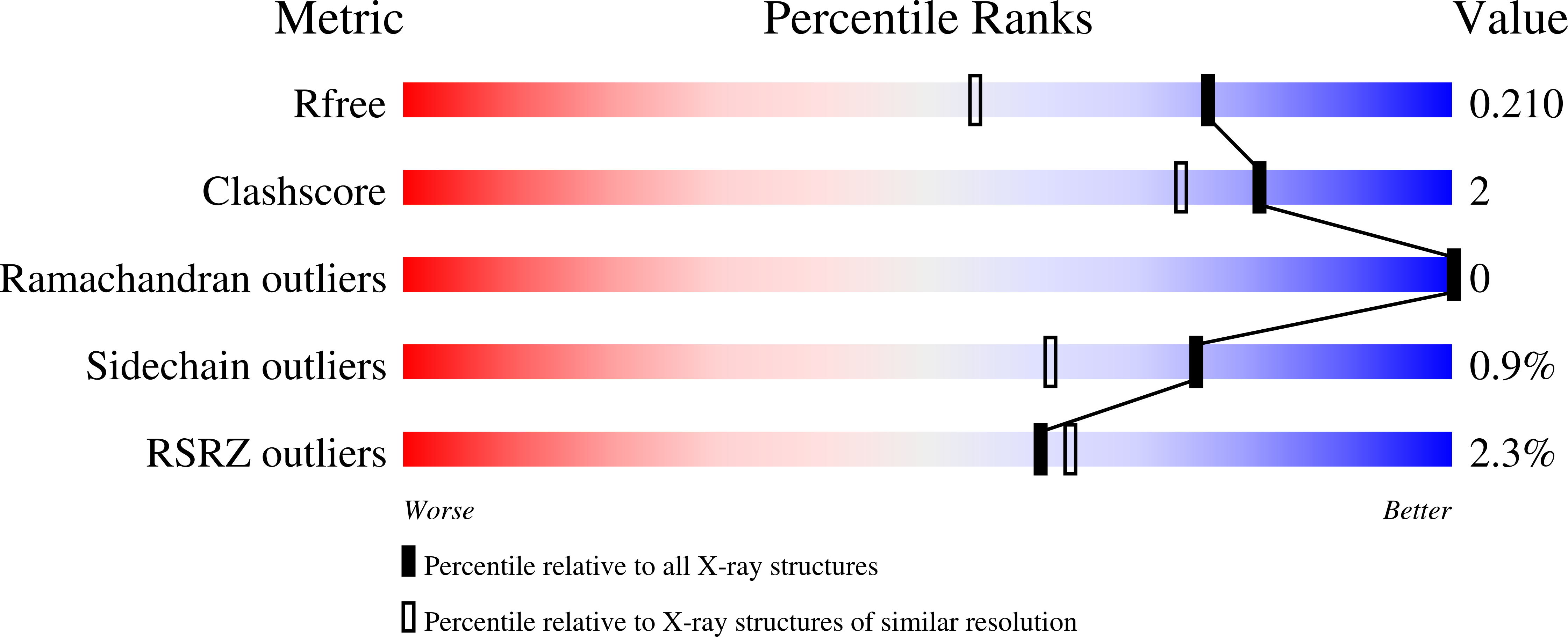
Resolution:
1.60 Å
R-Value Free:
0.21
R-Value Work:
0.17
R-Value Observed:
0.17
Space Group:
P 21 21 21
Macromolecular Entities
Structures with similar UniProt ID
Protein Blast
Polymer Type:polypeptide(L)
Molecule:Glycosyltransferase
Gene (Uniprot):GGD45_005139, GXW80_17485
Chain IDs:A, B
Chain Length:279
Number of Molecules:2
Biological Source:Rhizobium tropici CIAT 899
Primary Citation
WrtF from Rhizobium tropici CIAT 899 is a GT-A fold fucosyltransferase that binds its donor nonproductively.
Proc.Natl.Acad.Sci.USA 122 e2512460122 e2512460122 (2025)
PMID: 41166418 DOI: 10.1073/pnas.2512460122

Abstact

l-Fucose, a 6-deoxy-monosaccharide, is often incorporated into O-antigens in Rhizobia where it serves as an important marker for host recognition. Here, we biochemically and structurally characterize WrtF, an O-antigen polysaccharide l-fucosyltransferase from the plant endosymbiont Rhizobium tropici CIAT 899. We show that WrtF transfers l-fucose from its donor, guanosine 5'-diphospho-β-l-fucose (GDP-Fuc), to a nonreducing end glucose, creating an α-(1→4) linkage. We determined structures of WrtF at resolutions between 1.45 and 2.3 Å in the apo state, and in complex with GDP, GDP-Fuc, nonacetylated and acetylated trisaccharide acceptors, and with a tetrasaccharide product. WrtF has an N-terminal glycosyltransferase A (GT-A) fold and a unique C-terminal α-helical bundle domain. The structures, combined with activity assays, and sequence analysis show that WrtF is unusual in using neither divalent cations nor basic residues to coordinate and stabilize the donor pyrophosphate. In contrast to almost all previously characterized GT-A glycosyltransferases that use α-linked donors that present the donor monosaccharide axially, GDP-Fuc presents its monosaccharide equatorially. GDP-Fuc binds in a compact conformation with the l-fucose endocyclic oxygen and C6 methyl packed against ribose C5 and C4, respectively. This binding mode paradoxically shields the anomeric carbon from attack; superposition of the donor and acceptor complexes suggests that these substrates would sterically hinder one another and do not appropriately orient to form a Michaelis complex. We therefore propose that the donor l-fucose must reorient in the active site prior to turnover. The preferred occluded donor binding mode may serve to "prescreen" GDP-Fuc from structurally similar candidate donors, while also minimizing counterproductive donor hydrolysis.

Legend

Protein

Chemical

Disease

Primary Citation of related structures
Feedback Form
Name
Email
Institute
Feedback