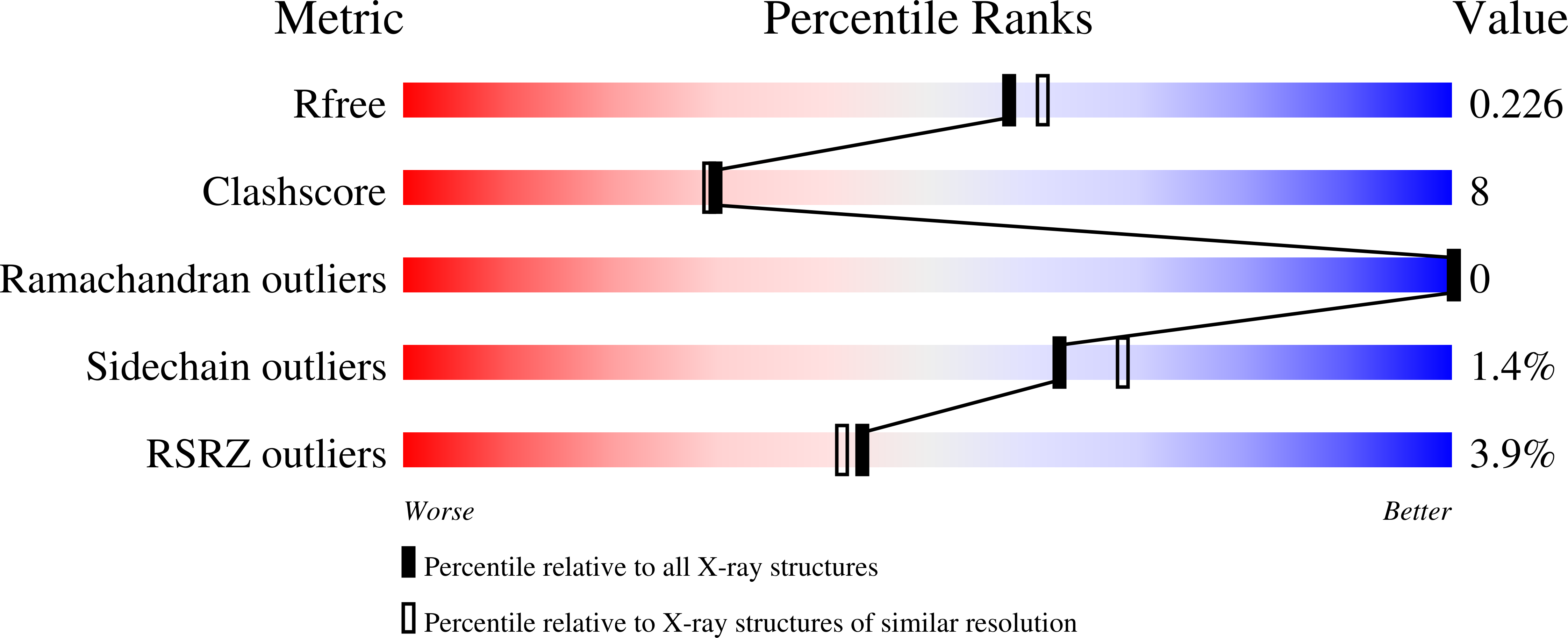
9OH7 image
Deposition Date 2025-05-02
Release Date 2025-07-23
Last Version Date 2025-07-23
Entry Detail
PDB ID:
9OH7
Keywords:
Title:
M13F/H117A/M121H Azurin with Cu(II), pH 7.4
Biological Source:
Source Organism:
Method Details:
Experimental Method:
Resolution:
2.00 Å
R-Value Free:
0.26
R-Value Work:
0.21
R-Value Observed:
0.21
Space Group:
P 1 21 1
Macromolecular Entities
Structures with similar UniProt ID
Protein Blast
Polymer Type:polypeptide(L)
Molecule:Azurin
Gene (Uniprot):azu
Mutagens:M13F, H117A, M121H
Chain IDs:A (auth: B), B (auth: A), C (auth: E), D (auth: G)
Chain Length:128
Number of Molecules:4
Biological Source:Pseudomonas aeruginosa PAO1
Primary Citation
Beyond Blue: Systematic Modulation of Electronic Structure and Redox Properties of Type 1 Copper in Azurin.
J.Am.Chem.Soc. 147 24825 24837 (2025)
PMID: 40626760 DOI: 10.1021/jacs.5c07009

Abstact

The reduction potentials of metal ions (E°'), crucial for optimizing biological processes like electron transfer and catalysis, are finely tuned by interactions between the primary and secondary coordination spheres (PCS, SCS). While previous successes in tuning E°' in azurin have provided deeper insights into how the SCS influences electronic structure and associated redox properties of "classic" blue copper proteins, our understanding of E°' tuning in other subclasses of type 1 Cu (T1Cu) proteins, such as green and red copper proteins, remains rudimentary. To address this issue, we report the design of a green copper center in azurin where an equatorial-to-axial shift in a histidine binding interaction leads to reorientation of the Cu-centered redox active molecular orbital and a +100 mV shift in E°'. In contrast to a 22 mV decrease in E°' when a hydrophobic interaction is introduced in wild-type azurin through the Met13Phe mutation, this same mutation leads to a 65 mV increase in our designed green Cu azurin. More importantly, using a combination of EPR spectroscopy, protein crystallography, and quantum mechanical calculations, we uncover correlations between E°', d-s orbital mixing, and the angle between SCys-Cu and NδH46-Cu bonds, ∠(SCys-Cu-NδH46), allowing rationalization of increases in E°' of green Cu proteins through an entropically driven T-shape distortion. By providing direct connections between geometry, electronic structure, and functional properties such as E°', this work opens previously unexplored routes to systematically modulating E°' through the combination of spatial reorientation of the redox active molecular orbital and varying geometric distortion in the primary coordination sphere.

Legend

Protein

Chemical

Disease

Primary Citation of related structures