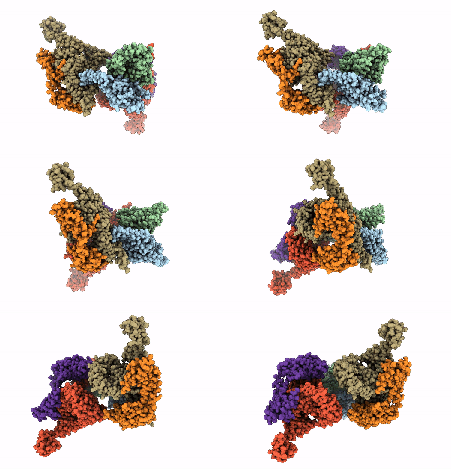
9OG0 image
Deposition Date 2025-04-30
Release Date 2025-07-30
Last Version Date 2025-07-30
Entry Detail
PDB ID:
9OG0
Title:
Cryo-EM structure of OS9-SEL1L-HRD1 dimer
Biological Source:
Source Organism:
Homo sapiens (Taxon ID: 9606)
Mus musculus (Taxon ID: 10090)
Host Organism:
Method Details:
Experimental Method:
Resolution:
3.64 Å
Aggregation State:
PARTICLE
Reconstruction Method:
SINGLE PARTICLE
Macromolecular Entities
Structures with similar UniProt ID
Protein Blast
Polymer Type:polypeptide(L)
Molecule:E3 ubiquitin-protein ligase synoviolin
Gene (Uniprot):SYVN1
Chain IDs:A, B
Chain Length:617
Number of Molecules:2
Biological Source:Homo sapiens
Structures with similar UniProt ID
Protein Blast
Polymer Type:polypeptide(L)
Molecule:Protein sel-1 homolog 1
Gene (Uniprot):Sel1l
Chain IDs:C, D
Chain Length:790
Number of Molecules:2
Biological Source:Mus musculus
Structures with similar UniProt ID
Protein Blast
Polymer Type:polypeptide(L)
Molecule:Isoform 2 of Protein OS-9
Gene (Uniprot):OS9
Chain IDs:E, F
Chain Length:612
Number of Molecules:2
Biological Source:Homo sapiens
Ligand Molecules
Primary Citation
Structural basis and pathological implications of the dimeric OS9-SEL1L-HRD1 ERAD Core Complex.
Biorxiv ? ? ? (2025)
PMID: 40661598 DOI: 10.1101/2025.06.13.659592

Abstact

UNLABELLED The SEL1L-HRD1 complex represents the most conserved branch of endoplasmic reticulum (ER)-associated degradation (ERAD), a critical pathway that clears misfolded proteins to maintain ER proteostasis. However, the molecular organization and pathogenic mechanisms of mammalian ERAD have remained elusive. Here, we report the first cryo-EM structure of the core mammalian ERAD complex, comprising the ER lectin OS9, SEL1L, and the E3 ubiquitin ligase HRD1. The structure, validated by mutagenesis and crosslinking assays, reveals a dimeric assembly of the core complex in which SEL1L and OS9 form a claw-like configuration in the ER lumen that mediates substrate engagement, while HRD1 dimerizes within the membrane to facilitate substrate translocation. Pathogenic SEL1L mutations at the SEL1L-OS9 (Gly585Asp) and SEL1L-HRD1 (Ser658Pro) interfaces disrupt complex formation and impair ERAD activity. A newly identified disease-associated HRD1 variant (Ala91Asp), located in transmembrane helix 3, impairs HRD1 dimerization and substrate processing, underscoring the functional necessity of this interface and HRD1 dimerization. Finally, the structure also reveals two methionine-rich crevices flanking the HRD1 dimer, suggestive of substrate-conducting channels analogous to those in the ER membrane protein complex (EMC). These findings establish a structural framework for mammalian ERAD and elucidate how mutations destabilizing this machinery contribute to human disease. SUMMARY The dimeric structure of the human SEL1L-HRD1 ERAD core complex reveals key architectural and functional principles underlying the recognition and processing of misfolded proteins linked to human disease.

Legend

Protein

Chemical

Disease

Primary Citation of related structures