9ODJ image
Deposition Date 2025-04-27
Release Date 2026-01-07
Last Version Date 2026-01-07
Entry Detail
PDB ID:
9ODJ
Title:
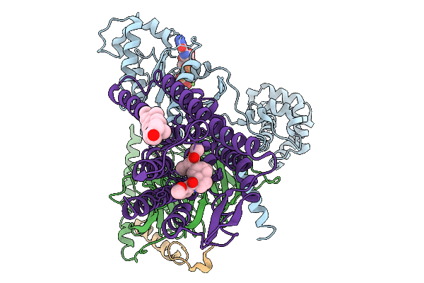
Structure of the MOR/Gi/Mitragynine Pseudoindoxil Complex, GTP-bound G-Primed, AHD 3DVA Sorted
Biological Source:
Source Organism:
Homo sapiens (Taxon ID: 9606)
Mus musculus (Taxon ID: 10090)
Host Organism:
Method Details:
Experimental Method:
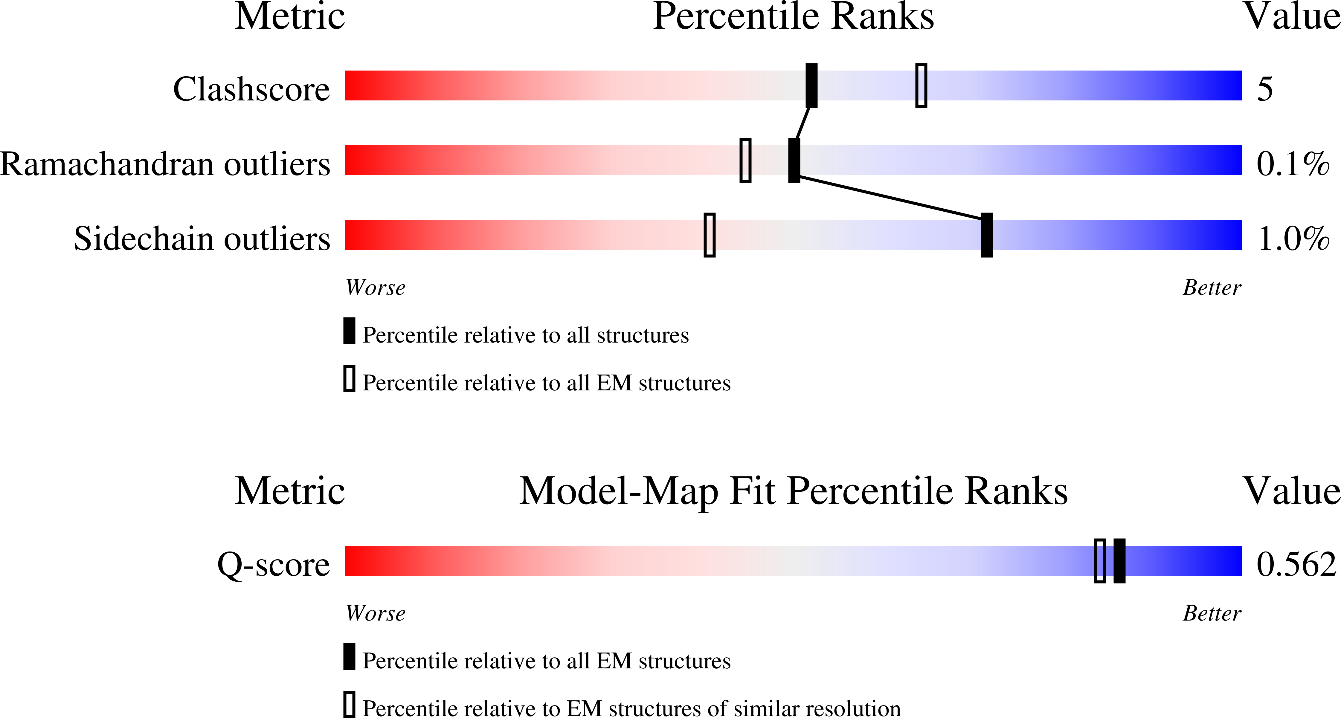
Resolution:
3.00 Å
Aggregation State:
PARTICLE
Reconstruction Method:
SINGLE PARTICLE
Macromolecular Entities
Structures with similar UniProt ID
Protein Blast
Polymer Type:polypeptide(L)
Molecule:Guanine nucleotide-binding protein G(i) subunit alpha-1
Gene (Uniprot):GNAI1
Chain IDs:D (auth: A)
Chain Length:354
Number of Molecules:1
Biological Source:Homo sapiens
Structures with similar UniProt ID
Protein Blast
Polymer Type:polypeptide(L)
Molecule:Guanine nucleotide-binding protein G(I)/G(S)/G(T) subunit beta-1
Gene (Uniprot):GNB1
Chain IDs:A (auth: B)
Chain Length:344
Number of Molecules:1
Biological Source:Homo sapiens
Structures with similar UniProt ID
Protein Blast
Polymer Type:polypeptide(L)
Molecule:Guanine nucleotide-binding protein G(I)/G(S)/G(O) subunit gamma-2
Gene (Uniprot):GNG2
Chain IDs:B (auth: C)
Chain Length:71
Number of Molecules:1
Biological Source:Homo sapiens
Structures with similar UniProt ID
Protein Blast
Polymer Type:polypeptide(L)
Molecule:Mu-type opioid receptor
Gene (Uniprot):Oprm1
Chain IDs:C (auth: D)
Chain Length:426
Number of Molecules:1
Biological Source:Mus musculus
Primary Citation
Non-equilibrium snapshots of ligand efficacy at the mu-opioid receptor.
Nature ? ? ? (2025)
PMID: 41430437 DOI: 10.1038/s41586-025-10056-4

Abstact

Distinct ligands for the same G-protein coupled receptor (GPCR) activate intracellular signaling partners to varying extents, but the molecular mechanisms driving these differences remain elusive. Hypothesizing that such differences in signaling efficacy may be captured structurally in intermediate states under non-equilibrium conditions, we implemented a time-resolved (TR) cryo-EM approach to visualize the GTP-induced activation of the Gαiβγ heterotrimer by the μ-opioid receptor (MOR) bound to three ligands displaying partial, full, or super-agonism on the receptor1. We resolved ensembles of conformational states along the G-protein activation pathway, including a previously unobserved intermediate state that enabled us to visualize receptor dynamics as a function of bound ligand. The results demonstrate ligand-dependent differences in state occupancy and conformational stability, with higher ligand efficacy correlating with increased dynamics of the receptor's transmembrane (TM) helices 5 and 6. Furthermore, we identify key mechanistic differences in the GTP-induced activation of Gi compared to Gs that likely underlie their distinct activation kinetics. Corroborated by molecular dynamics (MD) simulations and single-molecule fluorescence assays, these findings provide a dynamic structural landscape of GPCR-G-protein interactions for ligands of different efficacy and suggest partial agonists may produce a 'kinetic trap' during G-protein activation.

Legend

Protein

Chemical

Disease

Primary Citation of related structures
Feedback Form
Name
Email
Institute
Feedback