9OC4 image
Deposition Date 2025-04-23
Release Date 2025-05-21
Last Version Date 2025-07-30
Entry Detail
PDB ID:
9OC4
Title:
High-resolution cryo-EM structure of KdpFABC in the E1P-ADP state in lipid nanodisc
Biological Source:
Source Organism:
Host Organism:
Method Details:
Experimental Method:
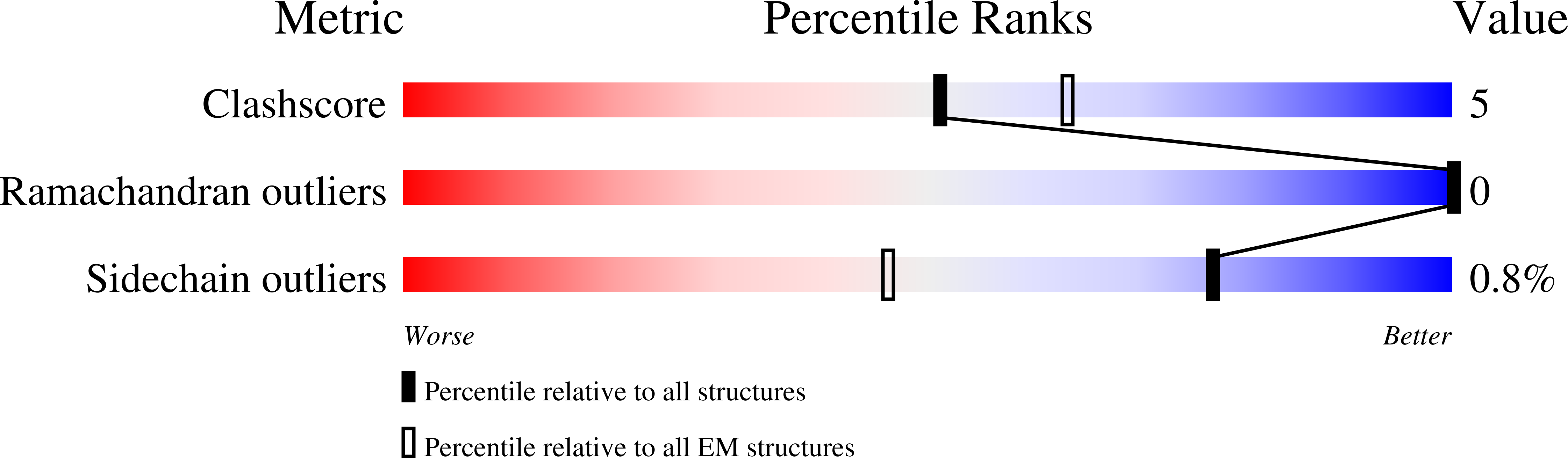
Resolution:
2.10 Å
Aggregation State:
PARTICLE
Reconstruction Method:
SINGLE PARTICLE
Macromolecular Entities
Structures with similar UniProt ID
Protein Blast
Polymer Type:polypeptide(L)
Molecule:Potassium-transporting ATPase potassium-binding subunit
Gene (Uniprot):kdpA
Chain IDs:A
Chain Length:557
Number of Molecules:1
Biological Source:Escherichia coli K-12
Structures with similar UniProt ID
Protein Blast
Polymer Type:polypeptide(L)
Molecule:Potassium-transporting ATPase ATP-binding subunit
Gene (Uniprot):kdpB
Chain IDs:B
Chain Length:682
Number of Molecules:1
Biological Source:Escherichia coli K-12
Structures with similar UniProt ID
Protein Blast
Polymer Type:polypeptide(L)
Molecule:Potassium-transporting ATPase KdpC subunit
Gene (Uniprot):kdpC
Chain IDs:C
Chain Length:208
Number of Molecules:1
Biological Source:Escherichia coli K-12
Structures with similar UniProt ID
Protein Blast
Polymer Type:polypeptide(L)
Molecule:Potassium-transporting ATPase KdpF subunit
Gene (Uniprot):kdpF
Chain IDs:D
Chain Length:29
Number of Molecules:1
Biological Source:Escherichia coli K-12
Modified Residue
Compound ID Chain ID Parent Comp ID Details 2D Image
PHD B ASP modified residue
Primary Citation
Conduction pathway for potassium through the E. coli pump KdpFABC.
Biorxiv ? ? ? (2025)
PMID: 40654642 DOI: 10.1101/2025.05.05.652293

Abstact

Under osmotic stress, bacteria express a heterotetrameric protein complex, KdpFABC, which functions as an ATP-dependent K+ pump to maintain intracellular potassium levels. The subunit KdpA belongs to the Superfamily of K+ Transporters and adopts a pseudo-tetrameric architecture with a membrane embedded selectivity filter as seen in K+ channels. KdpB belongs to the superfamily of P-type ATPases with a conserved binding site for ions within the membrane domain and three cytoplasmic domains that orchestrate ATP hydrolysis via an aspartyl phosphate intermediate. Previous work has hypothesized that K+ moves parallel to the membrane plane through a 40-Å long tunnel that connects the selectivity filter of KdpA with the binding site in KdpB. In the current work, we have reconstituted KdpFABC into lipid nanodiscs and used cryo-EM to image the wild-type pump under turnover conditions. We present a 2.1 Å structure of the E1~P·ADP conformation, which reveals new features of the conduction pathway. This map shows exceedingly strong densities within the selectivity filter and at the canonical binding site, consistent with K+ bound at each of these sites in this conformation. Many water molecules occupy a vestibule and the proximal end of the tunnel, which becomes markedly hydrophobic and dewetted at the subunit interface. We go on to use ATPase and ion transport assays to assess effects of numerous mutations along this proposed conduction pathway. The results confirm that K+ ions pass through the tunnel and support the existence of a low affinity site in KdpB for releasing these ions to the cytoplasm. Taken together, these data shed new light on the unique partnership between a transmembrane channel and an ATP-driven pump in maintaining the large electrochemical K+ gradient essential for bacterial survival.

Legend

Protein

Chemical

Disease

Primary Citation of related structures
Feedback Form
Name
Email
Institute
Feedback