9NWW image
Deposition Date 2025-03-24
Release Date 2025-12-17
Last Version Date 2025-12-17
Entry Detail
PDB ID:
9NWW
Keywords:
Title:
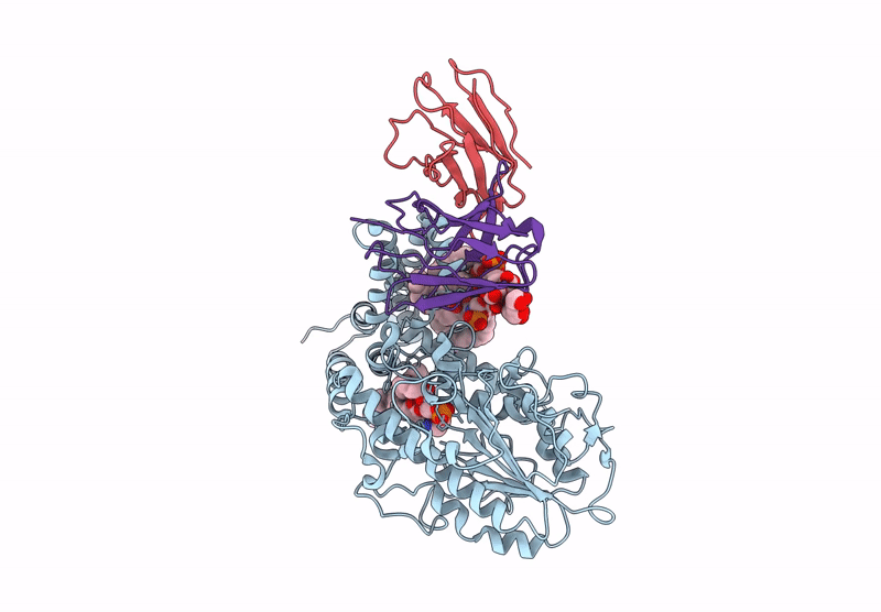
Single-particle cryo-EM structure of the first variant of mobilized colistin resistance (MCR-1) in its ligand-bound state
Biological Source:
Source Organism(s):
Escherichia coli (Taxon ID: 562)
Homo sapiens (Taxon ID: 9606)
Method Details:
Experimental Method:
Resolution:
3.58 Å
Aggregation State:
PARTICLE
Reconstruction Method:
SINGLE PARTICLE
Macromolecular Entities
Structures with similar UniProt ID
Protein Blast
Polymer Type:polypeptide(L)
Molecule:Probable phosphatidylethanolamine transferase Mcr-1
Gene (Uniprot):mcr1
Chain IDs:A (auth: B)
Chain Length:577
Number of Molecules:1
Biological Source:Escherichia coli
Polymer Type:polypeptide(L)
Molecule:Fab (MR6) Light (L) Chain
Chain IDs:B (auth: C)
Chain Length:214
Number of Molecules:1
Biological Source:Homo sapiens
Polymer Type:polypeptide(L)
Molecule:Fab (MR6) Heavy (H) Chain
Chain IDs:C (auth: D)
Chain Length:235
Number of Molecules:1
Biological Source:Homo sapiens
Primary Citation
Mechanistic basis of antimicrobial resistance mediated by the phosphoethanolamine transferase MCR-1.
Nat Commun 16 10516 10516 (2025)
PMID: 41298376 DOI: 10.1038/s41467-025-65515-3

Abstact

Polymyxins are used to treat infections caused by multidrug-resistant Gram-negative bacteria. They are cationic peptides that target the negatively charged lipid A component of lipopolysaccharides, disrupting the outer membrane and lysing the cell. Polymyxin resistance is conferred by inner-membrane enzymes, such as phosphoethanolamine transferases, which add positively charged phosphoethanolamine to lipid A. Here, we present the structure of MCR-1, a plasmid-encoded phosphoethanolamine transferase, in its liganded form. The phosphatidylethanolamine donor substrate is bound near the active site in the periplasmic domain, and lipid A is bound over 20 Å away, within the transmembrane region. Integrating structural, biochemical, and drug-resistance data with computational analyses, we propose a two-state model in which the periplasmic domain rotates to bring the active site to lipid A, near the preferential phosphate modification site for MCR-1. This enzymatic mechanism may be generally applicable to other phosphoform transferases with large, globular soluble domains.

Legend

Protein

Chemical

Disease

Primary Citation of related structures
Feedback Form
Name
Email
Institute
Feedback